Pleural effusion !
A pleural effusion is fluid collected between the lung and the chest wall; it is a sign of another disease, such as heart failure, infection or cancer.
Updated 2026-10-29
Structures involved
Tap one to see what it is and open it in 3D.
In biochemistry
In histology
In the clinical exam
- Chest palpation: expansion and tactile fremitus
- Chest percussion: resonance, dullness and hyperresonance
- Lung auscultation: normal breath sounds
- Voice sounds: bronchophony, egophony and whispered pectoriloquy
- The pleuropulmonary syndromes: the comparison table to know
- Pleural effusion syndrome
- Atelectasis syndrome
In physiology
Why it happens
- Edema and effusions: the four mechanisms
- Pulmonary thromboembolism: where it comes from and what it does
- Vascular changes in acute inflammation: vasodilation, leakage, exudate vs transudate
- Starling forces at the capillary
What it is
Abnormal collection of fluid in the pleural space, between the parietal and visceral pleura. It is a sign of an underlying disease rather than a diagnosis in itself.
Who gets it
Very common in hospital practice. Heart failure is the most frequent cause of transudates; pneumonia and cancer are leading causes of exudates. In Colombia and Latin America tuberculosis remains an important cause of lymphocytic exudates in young adults.
Causes
- Transudate: heart failure, cirrhosis, nephrotic syndrome, hypoalbuminaemia
- Exudate: pneumonia (parapneumonic, empyema), tuberculosis, cancer
- Pulmonary embolism
- Autoimmune disease (lupus, rheumatoid arthritis)
- Pancreatitis and subdiaphragmatic abscess
- Chylothorax from thoracic duct injury; haemothorax after trauma
Risk factors
- Heart, liver or kidney failure
- Pneumonia
- Cancer, especially lung and breast cancer and lymphoma
- Tuberculosis exposure
- Chest trauma or surgery
Pathophysiology
Fluid forms when filtration into the pleural space exceeds lymphatic drainage. Transudates result from raised hydrostatic or reduced oncotic pressure with intact pleura; exudates result from inflammation, infection or tumour that increases capillary permeability or blocks lymphatics.
Classification
- Transudate vs exudate (Light's criteria)
- Parapneumonic: simple, complicated, empyema
- Malignant
- Special types: haemothorax, chylothorax
Symptoms and signs
- Breathlessness, depending on size and speed
- Pleuritic chest pain (in inflammatory causes)
- Dry cough
- Dullness to percussion over the fluid
- Reduced breath sounds and tactile fremitus
- Signs of the cause: fever, oedema, weight loss
Diagnosis
- Clinical
- Dullness and reduced breath sounds at the base; history points to the cause. Bilateral effusions with heart failure often need no tap.
- Tests
- Diagnostic thoracentesis: protein and LDH compared with serum (Light's criteria: an exudate meets at least one), pH, glucose, cell count, Gram stain and culture, cytology, and adenosine deaminase when TB is suspected.
- Imaging
- Chest X-ray shows blunting of the costodiaphragmatic angle or a meniscus. Ultrasound detects small volumes, shows septations and guides the tap; CT looks for the cause.
Differential diagnosis
- Lobar collapse or consolidation
- Pleural thickening
- Raised hemidiaphragm
- Pleural tumour (mesothelioma)
Goal of treatment
Find and treat the cause, relieve breathlessness and drain infected or rapidly reaccumulating fluid to prevent a trapped lung.
Treatment
- Medical
- Treatment of the cause: diuretics for heart failure, antibiotics for parapneumonic effusion, antituberculosis therapy for pleural TB.
- Procedure
- Therapeutic thoracentesis to relieve breathlessness from large effusions.
- Procedure
- Chest drain for empyema or complicated parapneumonic effusion; intrapleural fibrinolytic therapy in selected cases.
- Surgical
- Recurrent malignant effusion: pleurodesis or an indwelling pleural catheter; surgical decortication for organised empyema.
Complications
- Empyema
- Trapped lung and pleural fibrosis
- Re-expansion pulmonary oedema after large drainage
- Pneumothorax from thoracentesis
Prognosis
Depends on the cause: transudates improve with treatment of heart, liver or kidney disease; malignant effusion indicates advanced cancer with limited survival.
Test yourself
Think of the answer, then tap the question to see it.
What do Light's criteria compare to decide that a pleural effusion is an exudate?
Pleural fluid protein and LDH with serum values; meeting any one criterion classifies it as an exudate.
What is the most common cause of a transudative pleural effusion?
Congestive heart failure.
Which pleural fluid finding supports tuberculous pleurisy?
A lymphocyte-predominant exudate with raised adenosine deaminase (ADA).
Which pleural fluid findings suggest a complicated parapneumonic effusion needing drainage?
Frank pus, a positive Gram stain or culture, or low pH and low glucose.
Related conditions
- Community-acquired pneumonia
- Pulmonary tuberculosis
- Heart failure
- Pulmonary embolism
- Amoebic liver abscess
- Pneumothorax
- Lung cancer
- Haemothorax
Educational information, not medical advice. For any symptom, see a health professional.
Sources: Pleural Effusion (StatPearls (NCBI Bookshelf), 2024).
Spotted a mistake?We are always improving for the community. Tell us what is wrong and our team will review it.Report a mistake
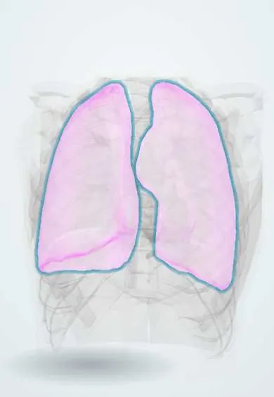
Representative model, for educational purposes only.