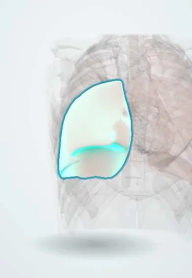
Atelectasis !
Atelectasis is the collapse of part or all of a lung, so that area cannot exchange air. It is common after surgery.
Updated 2026-11-01
Structures involved
Tap one to see what it is and open it in 3D.
- Right lower lobe
- Left lower lobe
- Lobar bronchi
- Main bronchi
- Segmental bronchi
- Pleura
- Diaphragm
- Trachea
- Lung fissures
In embryology
In the clinical exam
Why it happens
- Surface tension, surfactant and Laplace's law
- Lung and chest wall compliance
- Ventilation–perfusion matching and West zones
What it is
Incomplete expansion or collapse of alveoli in a segment, lobe or entire lung, with loss of volume. It is a finding with many causes rather than a single disease, and the most common pulmonary complication after surgery.
Who gets it
Develops in most patients under general anaesthesia and is a leading cause of hypoxaemia in the first days after abdominal and thoracic surgery; it is often blamed for early postoperative fever, but studies do not show that it causes fever. Also frequent in intensive care, in bedridden older adults and in children with foreign-body aspiration.
Causes
- Obstructive (resorptive): mucus plug, foreign body, tumour or enlarged lymph node blocking a bronchus
- Compressive: pleural effusion, pneumothorax, large tumour or raised diaphragm
- Passive or hypoventilation: shallow breathing from pain, sedation, anaesthesia or neuromuscular weakness
- Adhesive: surfactant deficiency (neonatal respiratory distress syndrome, ARDS)
- Cicatricial: scarring from tuberculosis or fibrosis
Risk factors
- General anaesthesia and upper abdominal or thoracic surgery
- Postoperative pain, opioids and immobility
- Obesity and pregnancy
- Smoking and chronic lung disease
- Rib fractures, neuromuscular disease, mechanical ventilation
Pathophysiology
When a bronchus is blocked, gas beyond it is absorbed into the blood and the alveoli collapse; breathing high oxygen concentrations speeds this up. Compression or shallow breathing without sighs also closes small airways. Collapsed but perfused alveoli create a shunt that causes hypoxaemia, and retained secretions in the collapsed area favour pneumonia.
Classification
- Obstructive (resorptive) and non-obstructive (compressive, passive, adhesive, cicatricial)
- Extent: subsegmental (plate-like), segmental, lobar or whole lung
- Acute (e.g. postoperative) or chronic (e.g. middle lobe syndrome)
Symptoms and signs
- Often no symptoms when small
- Shortness of breath and fast breathing
- Low oxygen saturation
- Low-grade fever may coexist after surgery, but it is not a reliable sign of atelectasis
- Reduced breath sounds and dullness over the affected area
- Tracheal deviation toward the affected side in large collapse
Diagnosis
- Clinical
- Suspected in a postoperative or bedridden patient with low saturation, fast breathing or fever, and reduced breath sounds at the bases. Nursing assessment: breath sounds, saturation, pain level and ability to cough deeply.
- Tests
- Pulse oximetry and arterial blood gas if needed. Bronchoscopy when an obstruction is suspected (foreign body, mucus plug that does not clear, tumour).
- Imaging
- Chest X-ray: opacity with signs of volume loss (displaced fissure, raised hemidiaphragm, tracheal and mediastinal shift toward the collapse). Lung ultrasound at the bedside; CT to look for an obstructing lesion.
Differential diagnosis
- Pneumonia (consolidation without volume loss)
- Pleural effusion (shift away from the opacity)
- Pulmonary embolism
- Pneumothorax
- Pulmonary oedema
Goal of treatment
Re-expand the collapsed lung, restore oxygenation and remove or treat the cause.
Treatment
- Rehabilitation
- Incentive spirometry, deep breathing and coughing exercises, early mobilisation and sitting upright; this is a core nursing intervention after surgery.
- Conservative
- Adequate pain control that allows deep breathing, chest physiotherapy, airway suctioning and humidified oxygen when needed.
- Procedure
- Positive airway pressure (CPAP) in selected patients; bronchoscopy to remove a mucus plug or foreign body.
- Medical
- Treatment of the cause: drainage of a pleural effusion or pneumothorax, antibiotics if pneumonia develops, management of a tumour.
Complications
- Hypoxaemia and respiratory failure
- Pneumonia in the collapsed segment
- Bronchiectasis in chronic collapse
- Longer hospital stay
Prognosis
Postoperative and mucus-plug atelectasis usually resolves within days with breathing exercises and mobilisation. Prognosis in obstructive atelectasis depends on the cause, especially when it is a tumour.
Prevention
- Stopping smoking before surgery
- Preoperative teaching and postoperative use of incentive spirometry
- Early mobilisation and good pain control
- Frequent position changes in bedridden patients
Test yourself
Think of the answer, then tap the question to see it.
What is the most common cause of low oxygen saturation in the first 48 hours after surgery, and does it explain fever?
Atelectasis from shallow breathing, pain and anaesthesia. It is often blamed for early fever, but the evidence does not support it as a cause, so a febrile patient still needs a search for other causes.
Toward which side does the trachea shift in large obstructive atelectasis?
Toward the collapsed side, because of volume loss (in contrast to a large effusion or tension pneumothorax).
Which nursing intervention is key to prevent postoperative atelectasis?
Incentive spirometry with deep breathing and coughing, plus early mobilisation and adequate pain control.
Why does breathing high-concentration oxygen favour collapse behind a blocked bronchus?
Oxygen is absorbed into the blood faster than nitrogen, so the trapped alveolar gas disappears quickly (absorption atelectasis).
Related conditions
- Acute respiratory failure (hypoxaemic and hypercapnic)
- Pneumothorax
- Pleural effusion
- Community-acquired pneumonia
- Hospital-acquired and ventilator-associated pneumonia
- Foreign body aspiration in children
- Lung cancer
- Pulmonary embolism
- Rib fractures
- Obesity
Educational information, not medical advice. For any symptom, see a health professional.
Sources: Atelectasis (MedlinePlus Medical Encyclopedia (U.S. National Library of Medicine), 2024).
Spotted a mistake?We are always improving for the community. Tell us what is wrong and our team will review it.Report a mistake
Representative model, for educational purposes only.