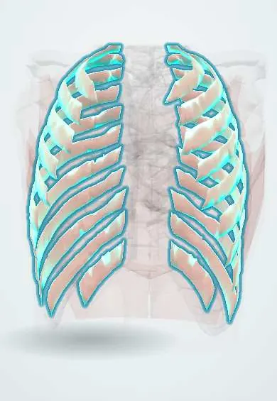
External intercostals !
The external intercostals are the outer layer of muscles between the ribs; they lift the ribs to help breathe in, especially during effort.
Official name: External intercostal muscles
Opens in the Durgical app, no sign-up needed.
What it is
Muscles between the ribs with fibres running down and forward.
What it does
Lift the ribs to help breathe in, especially when breathing hard.
Key facts
- Origin
- Inferior border of rib above
- Insertion
- Superior border of rib below; fibers run down and forward; replaced in front by external intercostal membrane
- Action
- Elevate ribs in inspiration; support intercostal space
- Nerve
- Intercostal nerves (T1–T11)
- Blood supply
- Posterior and anterior intercostal arteries
Relations
- Superficial
- Pectoralis major and minor, serratus anterior, latissimus dorsi and the back muscles, depending on the level
- Deep
- Internal intercostals, whose fibres cross theirs at right angles; the intercostal neurovascular bundle lies deeper still, under the internal layer
- Above
- Lower border of the rib above, from which they arise
- Below
- Upper border of the rib below, where they insert
How to find it
Not felt as a separate muscle: the intercostal spaces are palpated between the ribs, best on the lateral chest wall. On ultrasound with the probe across two ribs, the three muscle layers lie between the rib shadows, with the bright pleural line beneath.
Examination
- Chest expansion: hands on the lower ribs with the thumbs meeting at the midline; reduced or asymmetric movement on deep inspiration points to a problem on that side of the wall, pleura or lung
- Inspection for intercostal retractions: the spaces drawing in on inspiration signal increased work of breathing, a sign seen early in children
On imaging
On CT and MRI the three intercostal layers look like one thin muscle band between the ribs. Ultrasound with a linear probe across a space shows the layers between the rib shadows, above the bright pleural line: the view used to guide chest-wall blocks. After trauma or thoracotomy, CT shows lung herniating through a torn intercostal space.
Clinical notes
- Every needle or drain through the chest wall crosses all three intercostal layers, then the endothoracic fascia and the parietal pleura
- Paralysis of the intercostal muscles (high spinal cord injury) leaves the diaphragm to do the breathing, and the chest wall may draw in on inspiration
Development
Like the other intercostal layers, they come from the ventral extensions of the thoracic myotomes that grow round with the ribs; in the chest these stay separate segments, each with its own intercostal nerve, while over the abdomen they fuse into the flat muscle sheets.
Memory aid
External intercostals = hands in the front pockets (down and forward); internal intercostals run at right angles to them.
Related conditions
Test yourself
Think of the answer, then tap the question to see it.
In which direction do the fibres of the external intercostal muscles run?
Down and forward, like hands in the front pockets: the same direction as the external oblique.
What is the action of the external intercostal muscles?
They raise the ribs in inspiration: the upper ribs and sternum (pump-handle, more front-to-back depth) and the lower ribs (bucket-handle, more width).
What replaces the external intercostal muscles in front, between the costal cartilages?
The external intercostal membrane; the muscles run from the rib tubercles behind to the costochondral junctions in front.
What nerves supply the external intercostal muscles?
The intercostal nerves (T1–T11).
Educational information, not medical advice. For any symptom, see a health professional.
Its names
- Official Latin (TA2)
- musculi intercostales externi TA2 2311
- Official Spanish (DeCS)
- Músculos intercostales externos DeCS D007366
- English (TA2)
- External intercostal muscles
Related
Sources: Terminologia Anatomica (TA2); DeCS; Anatomy, Thorax, Wall Movements; Anatomy, Thoracotomy and the Collateral Intercostal Neurovascular Bundle; Anatomy of the Human Body (20th ed.): Development of the Muscles (Lea & Febiger (Gray's Anatomy, 1918), 1918) · adapted, Public domain.
Spotted a mistake?We are always improving for the community. Tell us what is wrong and our team will review it.Report a mistake
Representative model, for educational purposes only.