Conditions · Nephrology and urology
Renal cell carcinoma !
Renal cell carcinoma is the most common kidney cancer in adults. It is often found by chance on a scan and is curable by surgery when caught early.
Updated 2026-11-01
Structures involved
Tap one to see what it is and open it in 3D.
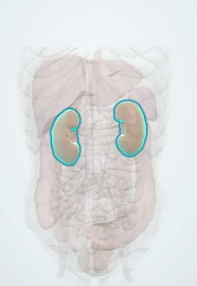
- Kidneys
- Left renal vein
- Right renal vein
- Inferior vena cava
- Left adrenal gland
- Right adrenal gland
- Renal pelvis
- Left renal artery
- Right renal artery
In the clinical exam
Why it happens
- Tumour suppressor genes: RB, TP53, APC, BRCA and the two-hit hypothesis
- Hereditary cancer syndromes: the table to know
- Tumour angiogenesis
- Paraneoplastic syndromes: the table to know
- Pathways of spread and the usual metastatic sites
- The hallmarks of cancer
Show 3 more
What it is
A malignant tumour arising from the epithelium of the renal tubules, most often the proximal tubule. It is the most common kidney cancer in adults.
Who gets it
Mostly affects adults aged 60–70, about twice as often men as women. Incidence is rising worldwide, partly because more tumours are found incidentally on ultrasound or CT. In Latin America it is less frequent than prostate or bladder cancer but follows the same risk pattern of smoking, obesity and hypertension.
Causes
- Loss of the VHL tumour suppressor gene (chromosome 3p) in most clear cell tumours
- Hereditary syndromes: von Hippel–Lindau disease, hereditary papillary renal cancer, Birt–Hogg–Dubé syndrome, tuberous sclerosis
Risk factors
- Smoking, the most important modifiable risk factor
- Obesity
- Hypertension
- Long-term dialysis with acquired cystic kidney disease
- Long-term misuse of some analgesics
- Family history and inherited syndromes
- Male sex and older age
Pathophysiology
In clear cell carcinoma, loss of VHL stabilises hypoxia-inducible factors, which switch on growth signals such as VEGF; this makes the tumour very vascular. It tends to grow into the renal vein and inferior vena cava and spreads by blood to lung, bone, liver and brain. Tumour products can cause paraneoplastic effects: erythropoietin (polycythaemia), PTH-related peptide (hypercalcaemia), renin (hypertension) and others.
Classification
- Clear cell carcinoma (most common, linked to VHL)
- Papillary carcinoma (types associated with MET or FH changes)
- Chromophobe carcinoma (better prognosis)
- Collecting duct and medullary carcinoma (rare, aggressive; medullary associated with sickle cell trait)
- Staging by TNM: I–II confined to the kidney, III into major veins or nodes, IV beyond Gerota fascia or distant spread
Symptoms and signs
- Often no symptoms; found incidentally on imaging
- Haematuria (microscopic or visible)
- Flank pain
- Palpable abdominal or flank mass (the classic triad with haematuria and pain is seen in a minority)
- Weight loss, fever, fatigue and anaemia
- A left varicocele that does not empty when lying down (left renal vein obstruction)
Diagnosis
- Clinical
- History of smoking and other risk factors, abdominal and flank examination, check for varicocele and leg oedema.
- Tests
- Urinalysis for haematuria, blood count (anaemia or polycythaemia), creatinine, calcium, liver function and lactate dehydrogenase. Biopsy is often unnecessary when imaging is typical; it is used for small masses, before ablation or before systemic therapy.
- Imaging
- Contrast-enhanced CT of the abdomen is the key test, showing an enhancing solid mass; ultrasound separates solid masses from simple cysts; MRI when contrast is contraindicated or to assess venous thrombus. Chest CT (and bone scan or brain imaging if symptomatic) for staging.
Differential diagnosis
- Simple or complex renal cyst (Bosniak classification)
- Angiomyolipoma (fat-containing benign tumour)
- Oncocytoma
- Urothelial carcinoma of the renal pelvis
- Renal abscess or xanthogranulomatous pyelonephritis
- Metastasis or lymphoma in the kidney
Goal of treatment
Cure localised disease while preserving as much kidney function as possible; in advanced disease, control the cancer and symptoms and prolong life.
Treatment
- Surgical
- Partial nephrectomy for small tumours to save kidney tissue; radical nephrectomy (kidney, surrounding fat and sometimes the adrenal gland and nodes) for larger tumours or venous extension.
- Procedure
- Thermal ablation (radiofrequency, microwave or cryoablation) for small tumours in people who are poor surgical candidates; arterial embolisation is used mainly to control bleeding or symptoms, not as cure.
- Follow-up
- Active surveillance of small masses in older or frail patients, and imaging follow-up after surgery.
- Medical
- Advanced disease is treated with immune checkpoint inhibitors and targeted therapies such as VEGF-receptor tyrosine kinase inhibitors and mTOR inhibitors; conventional chemotherapy is largely ineffective.
- Procedure
- Radiotherapy, mainly stereotactic, for painful bone or brain metastases.
Complications
- Metastasis to lung ("cannonball" nodules), bone, liver and brain
- Tumour thrombus in the renal vein and inferior vena cava
- Paraneoplastic syndromes: polycythaemia, hypercalcaemia, hypertension, Stauffer syndrome (liver dysfunction without metastasis)
- Chronic kidney disease after nephrectomy
Prognosis
Excellent for small tumours confined to the kidney after surgery; much worse with distant spread, although immunotherapy and targeted therapy have clearly improved survival. Late recurrences can occur, so follow-up is long.
Prevention
- Not smoking
- Maintaining a healthy weight and controlling blood pressure
- Surveillance imaging in people with von Hippel–Lindau disease and other hereditary syndromes
Test yourself
Think of the answer, then tap the question to see it.
Which gene is classically lost in clear cell renal cell carcinoma?
The VHL tumour suppressor gene on chromosome 3p, which stabilises hypoxia-inducible factor and drives angiogenesis.
What is the classic (but uncommon) triad of renal cell carcinoma?
Haematuria, flank pain and a palpable flank mass.
Why may a left varicocele suggest renal cell carcinoma?
The left testicular vein drains into the left renal vein; tumour invasion of that vein blocks drainage.
What are two paraneoplastic effects of renal cell carcinoma?
Polycythaemia from erythropoietin and hypercalcaemia from PTH-related peptide (also hypertension and Stauffer syndrome).
What is the main curative treatment for localised renal cell carcinoma?
Surgery: partial or radical nephrectomy.
Related conditions
- Autosomal dominant polycystic kidney disease
- Varicocele
- Chronic kidney disease
- Bladder cancer
- Wilms tumour (nephroblastoma)
- Kidney stones (urolithiasis)
- Hypercalcaemia
- Arterial hypertension
Educational information, not medical advice. For any symptom, see a health professional.
Sources: Renal cell carcinoma: MedlinePlus Medical Encyclopedia (MedlinePlus (U.S. National Library of Medicine), 2024); Renal Cell Cancer Treatment (PDQ®)–Patient Version (National Cancer Institute (NCI), 2025).
Spotted a mistake?We are always improving for the community. Tell us what is wrong and our team will review it.Report a mistake
Representative model, for educational purposes only.