Conditions · Nephrology and urology
Renovascular hypertension (renal artery stenosis) !
A narrowing of the artery to one or both kidneys. The kidney senses low flow and raises blood pressure, which can become hard to control.
Updated 2026-11-01
Structures involved
Tap one to see what it is and open it in 3D.
- Right renal artery
- Left renal artery
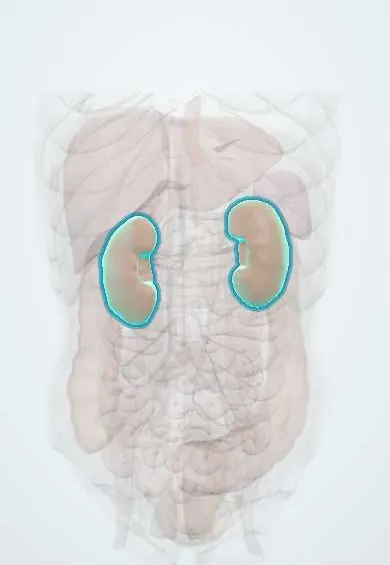
- Kidneys
- Abdominal aorta
- Vessels inside the kidney
- Right adrenal gland
- Left adrenal gland
In the clinical exam
Why it happens
- The renin–angiotensin–aldosterone system
- Long-term blood pressure control by the kidney
- Physiology of hypertension
- Renal blood flow, autoregulation and tubuloglomerular feedback
- Angiotensin-converting enzyme (ACE)
What it is
Narrowing of one or both renal arteries that reduces blood flow to the kidney. It is a potentially curable cause of secondary hypertension (kidney parenchymal disease is the most common secondary cause) and can also cause ischaemic kidney disease.
Who gets it
Atherosclerotic disease is the usual form in older adults with vascular risk factors; fibromuscular dysplasia mostly affects women under 50. It is an important cause to consider in Latin America given the high burden of hypertension, diabetes and smoking.
Causes
- Atherosclerosis at the origin of the renal artery (most cases, older adults)
- Fibromuscular dysplasia: a non-inflammatory wall disease with a "string of beads" appearance, in the middle or distal artery (young women)
- Less often: vasculitis such as Takayasu arteritis, dissection, external compression or thrombosis
Risk factors
- Older age, smoking, diabetes and dyslipidaemia
- Coronary, carotid or peripheral artery disease
- Female sex and younger age for fibromuscular dysplasia
- Heavy alcohol or cocaine use
Pathophysiology
Low pressure beyond the narrowing is sensed by the juxtaglomerular cells, which release renin. Angiotensin II constricts vessels and aldosterone retains sodium and water, so blood pressure rises (high-renin hypertension), often with low potassium. In bilateral disease or a single kidney, glomerular filtration depends on angiotensin II, which explains the fall in function with ACE inhibitors or ARBs.
Classification
- By cause: atherosclerotic or fibromuscular dysplasia
- Unilateral or bilateral (or stenosis of a solitary kidney)
- Haemodynamically significant or not
Symptoms and signs
- Often no symptoms apart from high blood pressure
- Hypertension starting before 30 (especially without family history) or after 55
- Resistant hypertension despite three drugs including a diuretic
- Sudden worsening of previously controlled blood pressure
- Abdominal or flank bruit
- Unexplained decline in kidney function or a small kidney on one side
Diagnosis
- Clinical
- Suspect it in hypertension with typical clues: age of onset, resistance, flash pulmonary oedema, abdominal bruit, kidney asymmetry or a fall in function after ACE inhibitors or ARBs.
- Tests
- Creatinine and eGFR, potassium (often low), urinalysis and urine albumin; plasma renin and aldosterone may be high. Renal function is rechecked after starting renin–angiotensin blockers.
- Imaging
- Doppler (duplex) ultrasound of the renal arteries as a first non-invasive test; CT or MR angiography. Catheter angiography is the reference standard and is done when intervention is planned.
Differential diagnosis
- Essential (primary) hypertension
- Primary hyperaldosteronism
- Chronic kidney parenchymal disease
- Pheochromocytoma
- Coarctation of the aorta
- Obstructive sleep apnoea or drug-induced hypertension
Goal of treatment
Control blood pressure, preserve kidney function and reduce cardiovascular risk; revascularise selected patients who benefit.
Treatment
- Lifestyle
- Stopping smoking, salt reduction, a heart-healthy diet, physical activity and limiting alcohol.
- Medical
- Antihypertensive drugs, often including ACE inhibitors or ARBs with close monitoring of creatinine and potassium (caution in bilateral disease or a single kidney); calcium-channel blockers and diuretics as needed.
- Medical
- Atherosclerotic disease: statins and, usually, antiplatelet therapy, plus control of diabetes.
- Procedure
- Angioplasty, with a stent for atherosclerotic lesions, is first choice in fibromuscular dysplasia and reserved in atherosclerosis for flash pulmonary oedema, refractory hypertension or progressive kidney loss.
- Follow-up
- Regular blood pressure, creatinine and potassium checks; imaging follow-up after revascularisation.
Complications
- Ischaemic nephropathy and chronic kidney disease
- Acute kidney injury with ACE inhibitors or ARBs in bilateral disease
- Flash pulmonary oedema and heart failure
- Stroke and myocardial infarction
- Hypertensive retinopathy
Prognosis
Fibromuscular dysplasia responds well to angioplasty and blood pressure often improves or normalises. In atherosclerotic disease, outcome is driven by overall cardiovascular disease; medical therapy is the mainstay because routine stenting has not shown better results than optimal drug treatment.
Prevention
- Control of atherosclerotic risk factors: not smoking, blood pressure, glucose and lipids
Test yourself
Think of the answer, then tap the question to see it.
What are the two main causes of renal artery stenosis and whom does each affect?
Atherosclerosis in older adults with vascular risk factors, and fibromuscular dysplasia in young women.
What is the classic angiographic appearance of fibromuscular dysplasia?
A "string of beads" in the middle or distal renal artery.
Why can creatinine rise sharply after an ACE inhibitor in bilateral stenosis?
Filtration depends on angiotensin II constricting the efferent arteriole; blocking it drops glomerular pressure.
Which non-invasive test is usually the first to screen for it?
Doppler (duplex) ultrasound of the renal arteries, with CT or MR angiography as alternatives.
Related conditions
- Arterial hypertension
- Chronic kidney disease
- Acute kidney injury
- Atherosclerosis and dyslipidaemia
- Peripheral artery disease
- Abdominal aortic aneurysm
- Primary hyperaldosteronism (Conn syndrome)
- Heart failure
- Hypokalaemia
Educational information, not medical advice. For any symptom, see a health professional.
Sources: Renal Artery Stenosis (NIDDK (National Institutes of Health), 2024); Renovascular hypertension: MedlinePlus Medical Encyclopedia (MedlinePlus (U.S. National Library of Medicine), 2024); Harrison's Principles of Internal Medicine, 21st ed., ch. 277 Hypertension (McGraw Hill, 2022).
Spotted a mistake?We are always improving for the community. Tell us what is wrong and our team will review it.Report a mistake
Representative model, for educational purposes only.