
Skin grafts and flaps: covering a wound
When a wound is too big to close, it is covered with transplanted skin (a graft) or with tissue that keeps its own blood supply (a flap).
What it is
A skin graft is skin cut completely free from a donor site and laid on a wound bed, which must grow new vessels into it to keep it alive. Split-thickness grafts carry the epidermis and part of the dermis; full-thickness grafts carry the whole dermis. A flap is tissue (skin, fat, fascia, muscle or bone) moved with its own blood supply, either still attached (pedicled) or reconnected to new vessels under the microscope (free flap).
Who gets it
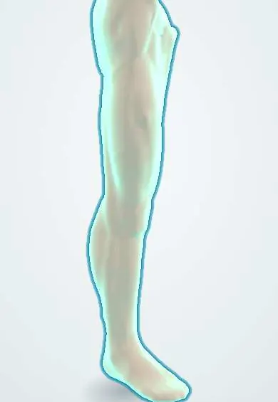
Skin grafting is one of the commonest reconstructive procedures, used above all for burns, traumatic skin loss, necrotising infection debridement, chronic leg and foot ulcers and skin cancer excision. Flaps are used for pressure injuries, open fractures of the leg, breast reconstruction and head and neck cancer. In Colombia, burns in children and adults, road traffic injuries with open leg fractures and pressure injuries in people with spinal cord injury make graft and flap cover a frequent need in burn units and plastic surgery services.
Causes
- Burns: deep partial-thickness and full-thickness burns that will not heal in time on their own
- Traumatic skin loss and degloving, and open fractures needing soft tissue cover
- Wounds left after debridement of necrotising soft tissue infection
- Excision of skin cancers (basal cell carcinoma, squamous cell carcinoma, melanoma) leaving a defect too large to close
- Chronic wounds: pressure injuries, venous and diabetic foot ulcers once the bed is prepared
- Reconstruction after cancer surgery (breast, head and neck) and release of burn scar contractures
Risk factors
- Graft failure: haematoma or seroma under the graft, shear or movement, infection, a poorly vascularised bed
- Beds that do not take a graft: exposed bone without periosteum, tendon without paratenon, cartilage without perichondrium, exposed implants, previously irradiated tissue
- Smoking, diabetes, peripheral arterial disease, malnutrition and corticosteroid use
- Flap failure: kinking, compression or thrombosis of the pedicle, tight dressings, low blood pressure, cold, haematoma
- Heavy bacterial load on the wound, particularly beta-haemolytic streptococci
Pathophysiology
Graft take follows the blood supply. A graft laid on a healthy bed first sticks by fibrin and drinks nutrients from the bed's fluid (plasmatic imbibition, first one to two days) → bed and graft capillaries line up and join (inosculation, from about day two or three) → new vessels grow into the graft (revascularisation, during the first week) → the graft turns pink and adheres. Anything between the graft and the bed (blood, serous fluid, pus) or any sliding of the graft breaks those first vascular connections → the graft dies. A flap does not need the bed: it lives on its pedicle → arterial blockage makes it pale and cold; venous blockage makes it swollen, purple and congested, and venous thrombosis is the commoner cause of free flap loss.
Classification
- Split-thickness skin graft: epidermis and part of the dermis; can be meshed to cover a larger area; donor site heals on its own from skin appendages
- Full-thickness skin graft: epidermis and full dermis; better colour, texture and less contraction; donor site must be closed directly
- Other grafts: composite grafts (skin with cartilage), dermal substitutes and cultured cells, mainly in burns
- Flaps by blood supply: random pattern (no named vessel, limited length) or axial (a named artery and vein)
- Flaps by movement: local (advancement, rotation, transposition), regional pedicled (for example latissimus dorsi or medial gastrocnemius) or free (microvascular anastomosis)
- Flaps by tissue: cutaneous, fasciocutaneous, muscle, myocutaneous, perforator or bone-containing flaps
- The reconstructive ladder: healing by secondary intention → direct closure → delayed closure → skin graft → local flap → regional flap → free flap; negative pressure wound therapy and tissue expansion are added steps
Symptoms and signs
- Healthy graft: adherent, pink and blanching from the end of the first week
- Failing graft: lifted by a dark fluid collection, grey, white or black patches, or sliding over the bed
- Infected graft: purulent exudate, offensive smell, graft melting away, spreading redness
- Arterial flap problem: pale, cool, flat flap with slow or absent capillary refill
- Venous flap problem: swollen, purple or blue, tense flap with brisk capillary refill and dark bleeding on pinprick
- Donor site of a split-thickness graft: painful, oozing raw area like a graze that heals over about two to three weeks
Diagnosis
- Clinical
- Decide whether the bed can accept a graft: healthy granulation, good blood supply, no exposed bone, tendon or hardware, low bacterial load. After surgery, inspect the graft for adherence and colour when the dressing is first taken down, and monitor flaps regularly for colour, temperature, turgor, capillary refill and Doppler signal.
- Tests
- Wound swab or tissue culture if infection is suspected; blood glucose control in diabetes; nutritional markers when healing is poor; full blood count before major reconstruction.
- Imaging
- Hand-held Doppler to locate perforator vessels and to monitor flaps. CT or MR angiography to map vessels before free flaps, and arterial duplex ultrasound when limb blood supply is in doubt. Plain X-ray or MRI if osteomyelitis under the wound is suspected.
Differential diagnosis
- Wound that will heal on its own with good dressings (no graft needed)
- Necrotising soft tissue infection still progressing (needs debridement first)
- Osteomyelitis under a non-healing wound
- Malignancy in a long-standing chronic wound (Marjolin ulcer)
- Arterial insufficiency causing a non-healing wound
Graft or flap: what does the wound need?
| Feature | Split-thickness graft | Full-thickness graft | Flap |
|---|---|---|---|
| What moves | Epidermis and part of the dermis | Epidermis and full dermis | Tissue with its own artery and vein |
| Needs from the bed | A well-vascularised bed | A well-vascularised bed, small area | Nothing: brings its own supply; covers bone, tendon, hardware |
| Contraction and look | Contracts more; thinner, colour mismatch | Contracts less; best colour and texture | Bulk and durable cover; may look bulky |
| Donor site | Heals on its own over weeks; can be reharvested | Closed directly (behind the ear, groin, upper eyelid) | A second wound, sometimes a functional loss |
| Typical use | Large burns, big leg wounds | Face, eyelids, fingers | Open fractures, pressure injuries, breast and head and neck reconstruction |
Goal of treatment
Achieve durable, functional and acceptable-looking cover with the simplest option that does the job well, prepared on a clean, well-vascularised bed, and protect it until it is established.
Treatment
- Procedure
- Prepare the bed: debride dead tissue, control infection, treat oedema and arterial disease, and use negative pressure wound therapy where it helps form granulation tissue or bridge to definitive cover.
- Surgical
- Split-thickness skin graft harvested with a dermatome, meshed if a large area must be covered, fixed with sutures, staples or tissue glue, and held still with a bolster or negative pressure dressing.
- Surgical
- Full-thickness skin graft for small defects where look and low contraction matter (face, fingers), with the donor site closed directly.
- Surgical
- Flaps when the bed cannot feed a graft or bulk and durability are needed: local flaps for small defects, pedicled regional flaps for pressure injuries or the chest, free flaps for complex or distant defects.
- Follow-up
- Flap observations at regular intervals in the first days: colour, temperature, turgor, capillary refill and Doppler. Keep the patient warm, well hydrated and with adequate blood pressure, and avoid pressure on the pedicle. Early return to theatre for a compromised flap gives the best chance of salvage.
- Conservative
- Protect the graft from shear and pressure until it adheres (splint over joints, elevation of limbs); moist dressing of the donor site; leeches are sometimes used for a venously congested flap when the vein cannot be revised, with antibiotic cover because of the bacteria they carry.
- Rehabilitation
- Scar care after healing: compression garments, massage, sun protection and moisturising; physiotherapy and splints to prevent contracture across joints.
Complications
- Partial or complete graft loss
- Flap failure, partial necrosis or venous congestion
- Infection of the graft or donor site
- Haematoma or seroma under the graft or flap
- Graft contracture, especially across joints, the neck and eyelids
- Hypertrophic scarring and colour mismatch
- Donor site pain, delayed healing or functional loss after muscle harvest
Common traps
- A graft can be laid on any clean wound: it needs a bed that can grow vessels into it; bare bone, tendon, cartilage or hardware will not feed it, and these need a flap
- A pale, cool flap and a purple, swollen flap mean the same problem: pale is arterial inflow failure, purple and congested is venous outflow failure, and the second is the commoner cause of free flap loss
- The simplest rung of the reconstructive ladder is always best: the right choice is the one that gives durable, functional cover; sometimes a flap first avoids repeated failed grafts
- Full-thickness grafts are better grafts, so they should be used for everything: they take less reliably and their donor sites are small and must be closed; large wounds need split-thickness grafts
- Graft loss is usually caused by rejection: the patient's own skin is not rejected; grafts fail mostly from haematoma or seroma under them, shear and infection
Prognosis
On a well-prepared bed most split-thickness grafts take almost completely; failures are usually local and can be regrafted. Free flaps succeed in the large majority of cases when monitored closely and when a failing flap is taken back to theatre early. Long-term results depend on scar care and prevention of contracture.
Prevention
- Prepare the bed well before grafting and treat infection first
- Stop smoking before elective reconstruction and control blood glucose
- Immobilise the graft and avoid shear in the first week
- Meticulous haemostasis under grafts and flaps
- Structured flap monitoring by trained staff in the first days
Test yourself
Think of the answer, then tap the question to see it.
Why does a split-thickness skin graft donor site heal on its own, but a full-thickness graft donor site has to be sutured closed?
A split-thickness harvest leaves the deep dermis with hair follicles and sweat glands, whose cells regrow the epidermis. A full-thickness harvest removes all of the dermis and those appendages, so nothing is left to resurface it.
After an open tibial fracture, the bone is exposed without periosteum. Why will a skin graft fail, and what covers it?
Bare bone cannot grow vessels into a graft, so the graft starves. It needs a flap with its own blood supply, such as a local muscle flap (for example medial gastrocnemius) or a free flap.
On day one after a free flap, the flap is swollen, purple and refills very fast. What has happened and what is done?
Venous congestion, most likely a venous thrombosis or compressed pedicle. Release any tight dressing and call the surgeon for urgent return to theatre to revise the vein.
What is the commonest reason a skin graft does not take, and why?
Fluid under the graft (haematoma or seroma), along with shear: it separates the graft from the bed and breaks the first vascular connections that keep it alive.
Connections
Educational information, not medical advice. For any symptom, see a health professional.
Sources: Split-Thickness Skin Grafts (StatPearls (NCBI Bookshelf), 2025); Full-Thickness Skin Grafts (StatPearls (NCBI Bookshelf), 2025); Wound Healing Phases (StatPearls (NCBI Bookshelf), 2023); Skin graft (MedlinePlus (U.S. National Library of Medicine), 2025).
Spotted a mistake?We are always improving for the community. Tell us what is wrong and our team will review it.Report a mistake
Representative model, for educational purposes only.