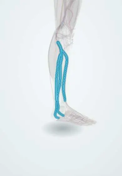
Venous leg ulcer !
A venous leg ulcer is a slow-healing wound of the lower leg, usually above the inner ankle, caused by high pressure in the leg veins.
Updated 2026-10-29
Structures involved
Tap one to see what it is and open it in 3D.
- Leg veins
- Perforating veins
- Great saphenous vein
- Small saphenous vein
- Tibia
- Crural fascia
- Soleus
- Gastrocnemius (inner head)
In histology
In the clinical exam
- Varicose veins: the Trendelenburg (Brodie–Trendelenburg) test
- Acute limb ischemia and chronic arterial insufficiency
Why it happens
- Abnormal repair: chronic wounds, hypertrophic scar, keloid, contracture
- What happens to a thrombus: propagation, embolism, dissolution, organization
- Healing of skin wounds: first and second intention
- What delays healing: infection, diabetes, nutrition, glucocorticoids, poor blood flow
- Granulation tissue and scar formation
- Starling forces at the capillary
What it is
A chronic wound of the lower leg caused by venous hypertension, typically in the gaiter area above the medial malleolus. It is the most common type of leg ulcer.
Who gets it
Most leg ulcers are venous; they become more common with age and tend to recur. They cause long periods of disability and large costs to health systems.
Causes
- Chronic venous insufficiency from valve failure
- Post-thrombotic syndrome after deep vein thrombosis
- Calf muscle pump failure (immobility, ankle stiffness)
- Minor trauma on fragile skin as a trigger
Risk factors
- Previous deep vein thrombosis
- Varicose veins
- Older age
- Obesity
- Reduced mobility
- Previous venous ulcer
Pathophysiology
Sustained venous hypertension leaks fluid and proteins into the tissues and draws in white cells that release inflammatory mediators. The skin becomes fibrotic and poorly oxygenated, so small injuries break down and fail to heal.
Classification
- CEAP class C6 (active) or C5 (healed)
- Pure venous or mixed arterial-venous, by the ankle-brachial index
Symptoms and signs
- Shallow ulcer with irregular edges and a moist, granulating base, often with heavy exudate
- Located in the gaiter area, most often above the medial malleolus
- Surrounding skin with haemosiderin pigmentation, eczema or lipodermatosclerosis
- Ankle swelling and aching that improves with elevation
Diagnosis
- Clinical
- Description of the wound and surrounding skin, pulses and the ankle-brachial index to rule out arterial disease before compression.
- Tests
- Swab culture only when clinical infection is present; blood glucose and other tests by context. Biopsy if the ulcer is atypical or does not heal.
- Imaging
- Venous duplex ultrasound to find reflux or obstruction that can be treated.
Differential diagnosis
- Arterial (ischaemic) ulcer
- Neuropathic (diabetic) ulcer
- Pyoderma gangrenosum
- Skin cancer (squamous cell or basal cell carcinoma, Marjolin ulcer)
- Vasculitic ulcer
- Cutaneous leishmaniasis in endemic areas
Goal of treatment
Heal the ulcer by reducing venous hypertension, then prevent recurrence.
Treatment
- Conservative
- Compression therapy (multilayer bandages or stockings), the cornerstone, only after arterial disease has been ruled out with the ankle-brachial index.
- Conservative
- Wound care: cleaning, debridement of dead tissue, dressings chosen by exudate, and care of the surrounding skin.
- Lifestyle
- Leg elevation, walking and ankle exercises.
- Medical
- Systemic antibiotics only for clinical infection; some guidelines include pentoxifylline or venoactive drugs to aid healing.
- Procedure
- Early ablation of superficial venous reflux speeds healing and lowers recurrence.
- Surgical
- Skin grafting for large or resistant ulcers.
Complications
- Cellulitis
- Recurrence
- Chronic pain and reduced quality of life
- Contact dermatitis from dressings
- Malignant change in a long-standing ulcer (rare)
Prognosis
Many ulcers heal with compression within months, but recurrence is common without long-term compression and treatment of venous reflux.
Prevention
- Lifelong compression after healing
- Treatment of superficial venous reflux
- Early treatment of deep vein thrombosis
- Skin care and weight control
Test yourself
Think of the answer, then tap the question to see it.
Where is a venous ulcer typically located?
In the gaiter area of the lower leg, most often above the medial malleolus.
What is the cornerstone of venous ulcer treatment?
Compression therapy, together with leg elevation and wound care.
What must be checked before applying compression to a leg ulcer?
The ankle-brachial index, to rule out significant arterial disease.
How does a venous ulcer differ from an arterial ulcer?
Venous: shallow, moist, irregular, near the medial malleolus. Arterial: deep, punched-out, painful, on the toes or foot.
Which malignancy can arise in a long-standing chronic ulcer?
Squamous cell carcinoma (Marjolin ulcer); a non-healing ulcer with atypical edges is biopsied.
Related conditions
- Chronic venous insufficiency
- Varicose veins
- Deep vein thrombosis
- Peripheral artery disease
- Diabetic foot
- Cellulitis of the leg
Educational information, not medical advice. For any symptom, see a health professional.
Sources: Venous Leg Ulcer (StatPearls (NCBI Bookshelf), 2026).
Spotted a mistake?We are always improving for the community. Tell us what is wrong and our team will review it.Report a mistake
Representative model, for educational purposes only.