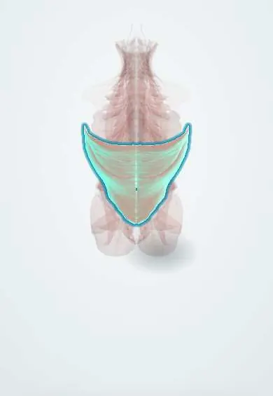
Latissimus dorsi !
The latissimus dorsi is the broad back muscle from the lower spine and pelvis to the upper arm; it pulls the arm down and back.
Official name: Latissimus dorsi muscle
Opens in the Durgical app, no sign-up needed.
What it is
The broad back muscle from the lower spine, thoracolumbar fascia and pelvis to the upper arm.
What it does
Pulls the arm down and back (climbing, rowing) and, through the fascia, helps stiffen the lower back.
Key facts
- Origin
- Spinous processes of T7–T12, thoracolumbar fascia, iliac crest, lower 3–4 ribs (often inferior angle of scapula)
- Insertion
- Floor of the intertubercular sulcus of the humerus
- Action
- Extends, adducts and medially rotates the humerus; raises the trunk when climbing
- Nerve
- Thoracodorsal nerve (C6–C8)
- Blood supply
- Thoracodorsal artery (from subscapular); posterior intercostal and lumbar perforators
Relations
- Superficial
- Skin and fat over most of it; trapezius overlaps its upper medial part
- Deep
- Erector spinae and serratus posterior inferior (through the thoracolumbar fascia), the lower ribs, the inferior angle of the scapula and teres major
- Lateral
- Its free border forms the posterior axillary fold with teres major; its rib slips interlock with the external oblique
- Below
- Iliac crest; the inferior lumbar triangle (of Petit) lies between its lateral border, the external oblique and the crest
How to find it
Grasping the posterior axillary fold between thumb and fingers holds it with teres major; the fold hardens when the person presses the arm down against resistance or coughs.
Examination
- Manual test: the person pulls the raised arm down and back against resistance, or coughs, while the examiner feels the posterior axillary fold; absent or weak contraction points to a thoracodorsal nerve lesion
On imaging
MRI shows it best: a broad, thin muscle wrapping the lower back and forming the posterior axillary fold with teres major. A tear of its tendon near the intertubercular groove of the humerus is rare and mostly seen in throwing athletes; MRI shows a retracted tendon with oedema. CT angiography can map the thoracodorsal artery before a flap, and ultrasound checks for a seroma at the donor site.
Clinical notes
- Latissimus dorsi flap on the thoracodorsal artery: a standard option to rebuild the breast and chest wall or to cover large defects
- Tendon transfer to restore external rotation in massive irreparable rotator cuff tears and in brachial plexus birth palsy
- A lumbar hernia can come through the inferior lumbar triangle at its lateral border
Anatomical variants
- Axillary arch (of Langer): a muscle slip from its edge crossing the axilla to pectoralis major, present in some people; it can press on axillary vessels or nerves and confuse axillary surgery
Development
A classic migrating muscle: it starts with the limb bud muscles in the lower neck and spreads down over the back, carrying its nerve with it. That is why a muscle of the lower back is supplied by the thoracodorsal nerve (C6–C8) from the brachial plexus.
Memory aid
The lady between two majors: latissimus dorsi inserts in the floor of the intertubercular sulcus, between pectoralis major (lateral lip) and teres major (medial lip).
Test yourself
Think of the answer, then tap the question to see it.
Where does the latissimus dorsi insert?
The floor of the intertubercular sulcus of the humerus, between pectoralis major and teres major («the lady between two majors»).
What are the action and nerve supply of the latissimus dorsi?
It extends, adducts and medially rotates the humerus; supplied by the thoracodorsal nerve (C6–C8).
How is the latissimus dorsi tested at the bedside?
Feeling the posterior axillary fold while the person pulls the raised arm down against resistance or coughs; weakness points to the thoracodorsal nerve.
Which triangle does the latissimus dorsi bound with the external oblique and iliac crest?
The inferior lumbar triangle (of Petit), a site of lumbar hernia.
On which vessel is a latissimus dorsi flap based?
The thoracodorsal artery; used to rebuild the breast and chest wall.
Educational information, not medical advice. For any symptom, see a health professional.
Its names
- Official Latin (TA2)
- musculus latissimus dorsi TA2 2231
- Official Spanish (DeCS)
- Músculo dorsal ancho DeCS D064171
- English (TA2)
- Latissimus dorsi muscle
Related
Sources: Terminologia Anatomica (TA2); DeCS; Anatomy, Back, Latissimus Dorsi; Anatomy of the Human Body (20th ed.): Development of the Muscles (Lea & Febiger (Gray's Anatomy, 1918), 1918) · adapted, Public domain.
Spotted a mistake?We are always improving for the community. Tell us what is wrong and our team will review it.Report a mistake
Representative model, for educational purposes only.