Osteomyelitis !
Osteomyelitis is an infection of bone, reaching it through the blood, from a nearby wound or ulcer, or after an injury or surgery.
Updated 2026-10-29
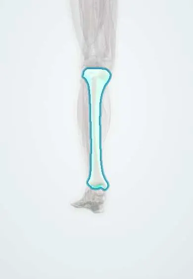
Structures involved
Tap one to see what it is and open it in 3D.
Microorganisms that cause it
In histology
Why it happens
- Outcomes of acute inflammation: resolution, abscess, scar or chronic inflammation
- Patterns of necrosis: coagulative, liquefactive, caseous, fat, fibrinoid, gangrenous
- Ossification, growth plate and bone remodeling
What it is
Infection of bone, which may be acute or chronic, and spreads through the blood, from nearby tissue (such as an ulcer) or directly after trauma or surgery.
Who gets it
In children it usually affects the long bones of the leg through the blood. In adults it is mostly linked to diabetic foot ulcers, open fractures and orthopaedic surgery, which are frequent problems in Colombia.
Causes
- Staphylococcus aureus (most common at all ages)
- Streptococci, Kingella kingae in young children
- Gram-negative bacteria and mixed flora in diabetic foot and after trauma
- Salmonella in sickle cell disease
- Mycobacterium tuberculosis in some cases
Risk factors
- Diabetes with foot ulcers
- Peripheral artery disease
- Open fractures and orthopaedic implants
- Sickle cell disease
- Intravenous drug use
- Immunosuppression
Pathophysiology
In children, bacteria in the blood lodge in the metaphysis, where slow-flowing looping vessels favour infection. Pus raises pressure, cuts off blood supply and leaves dead bone (sequestrum), surrounded by new bone (involucrum). In adults, infection more often spreads from a nearby ulcer or wound.
Classification
- Haematogenous versus contiguous spread or direct inoculation
- Acute versus chronic
- Cierny–Mader classification (anatomy of the infection and host status)
Symptoms and signs
- Bone pain, tenderness and limp or refusal to bear weight in children
- Fever and malaise in acute cases
- Swelling, warmth and redness over the bone
- Chronic draining sinus in chronic disease
- Ulcer with exposed or probe-reachable bone in diabetic foot
Diagnosis
- Clinical
- Localised bone tenderness, fever and, in the diabetic foot, a positive probe-to-bone test.
- Tests
- Blood count, ESR and CRP to support the diagnosis and follow response; blood cultures; bone biopsy for culture and histology is the reference standard.
- Imaging
- X-ray first, though changes appear only after about 2 weeks; MRI is the most sensitive test; bone scan or CT when MRI is not available.
Differential diagnosis
- Cellulitis
- Septic arthritis
- Charcot neuroarthropathy
- Bone tumours (Ewing sarcoma, osteosarcoma)
- Stress fracture
- Bone infarct in sickle cell disease
Goal of treatment
Eradicate the infection, remove dead bone, preserve function and stability, and prevent chronic disease.
Treatment
- Medical
- Prolonged antibiotics, started empirically and adjusted to cultures, often intravenous at first and then oral; duration criteria vary.
- Surgical
- Debridement of dead bone and abscess drainage; removal of infected implants; amputation in some diabetic feet.
- Procedure
- Bone biopsy, image-guided or surgical, to identify the organism before treatment when possible.
- Follow-up
- Clinical review and serial inflammatory markers to follow the response.
Drugs for this condition
- Cefalexin
- Cefazolin
- Ceftazidime
- Cloxacillin
- Dicloxacillin
- Oxacillin
- Nafcillin
- Flucloxacillin
- Vancomycin
- Imipenem–cilastatin
- Clindamycin
Complications
- Chronic osteomyelitis with sinus tracts
- Pathological fracture
- Septic arthritis
- Growth disturbance in children
- Amputation
- Squamous cell carcinoma in a long-standing sinus (Marjolin ulcer)
Prognosis
Acute haematogenous osteomyelitis in children usually heals with timely treatment. Chronic and diabetic foot osteomyelitis are harder to cure and often recur.
Prevention
- Good diabetic foot care
- Early treatment and careful cleaning of open fractures
- Antibiotic prophylaxis and sterile technique in orthopaedic surgery
Test yourself
Think of the answer, then tap the question to see it.
Where in the long bone does haematogenous osteomyelitis usually begin in children, and why?
In the metaphysis, where slow, looping blood vessels let bacteria settle.
What is the most common organism in osteomyelitis?
Staphylococcus aureus.
Which imaging test is most sensitive for early osteomyelitis?
MRI; plain X-ray changes usually take about 2 weeks to appear.
What is the reference standard to confirm osteomyelitis and identify the organism?
Bone biopsy with culture and histology.
What are a sequestrum and an involucrum?
A sequestrum is dead infected bone; an involucrum is new bone formed around it in chronic osteomyelitis.
Related conditions
- Diabetic foot
- Cellulitis of the leg
- Septic Arthritis of the Knee
- Tibial shaft fracture
- Vertebral osteomyelitis
Educational information, not medical advice. For any symptom, see a health professional.
Sources: Osteomyelitis (StatPearls (NCBI Bookshelf), 2023).
Spotted a mistake?We are always improving for the community. Tell us what is wrong and our team will review it.Report a mistake
Representative model, for educational purposes only.