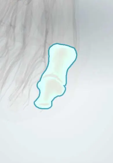
Diabetic foot !
Diabetic foot covers the ulcers, infections and tissue loss caused by diabetes through nerve damage and poor blood flow to the foot.
Updated 2026-10-29
Structures involved
Tap one to see what it is and open it in 3D.
- Big toe
- First metatarsal
- Calcaneus
- Dorsalis pedis artery
- Posterior tibial artery
- Tibial nerve
- Medial plantar nerve
- Fifth metatarsal
- Sesamoids
- Plantar fascia
In histology
- Wound healing: phases and scar
- Skin receptors: Meissner, Pacinian, Merkel, Ruffini and free nerve endings
Why it happens
- Abnormal repair: chronic wounds, hypertrophic scar, keloid, contracture
- What delays healing: infection, diabetes, nutrition, glucocorticoids, poor blood flow
- Healing of skin wounds: first and second intention
- Granulation tissue and scar formation
- Patterns of necrosis: coagulative, liquefactive, caseous, fat, fibrinoid, gangrenous
- Physiology of diabetes and ketoacidosis
What it is
Foot problems caused by diabetes: ulcers, infection and tissue loss that arise from the combination of nerve damage (neuropathy), poor blood flow (ischaemia) and infection.
Who gets it
A leading cause of non-traumatic lower-limb amputation worldwide. In Colombia and Latin America, the growing number of people with diabetes, late diagnosis and uneven access to foot care make it a major public health problem.
Causes
- Peripheral neuropathy: loss of protective sensation, foot deformity and dry skin
- Peripheral artery disease that reduces healing
- Repeated pressure or minor trauma (ill-fitting shoes, walking barefoot, burns)
- Bacterial infection entering through an ulcer
Risk factors
- Long-standing or poorly controlled diabetes
- Previous ulcer or amputation
- Smoking
- Foot deformity (claw toes, bunion, Charcot foot)
- Chronic kidney disease and diabetic retinopathy
- Poor footwear and limited access to foot care
Pathophysiology
High glucose damages nerves, so the patient stops feeling pressure and small injuries, and motor and autonomic loss deform the foot and dry the skin. Pressure points then break down into ulcers. Ischaemia slows healing, and impaired immunity lets bacteria spread to soft tissue and bone.
Classification
- Wagner classification (grades 0–5, from at-risk foot to gangrene of the whole foot)
- University of Texas classification (depth combined with infection and ischaemia)
- IWGDF/IDSA grading of infection severity (mild, moderate, severe)
- Neuropathic, ischaemic or neuroischaemic ulcer
Symptoms and signs
- Painless ulcer, often under the metatarsal heads, big toe or heel
- Numbness, tingling or burning in the feet
- Calluses, dry cracked skin and toe deformities
- Cold foot with weak or absent pulses when ischaemia is present
- Redness, warmth, swelling or discharge when infected
Diagnosis
- Clinical
- Inspection of both feet, 10 g monofilament test for protective sensation, vibration testing, palpation of pulses and probe-to-bone test for deep ulcers.
- Tests
- Glucose and HbA1c, blood count and inflammatory markers; deep tissue or bone culture rather than surface swabs; ankle–brachial index (may be falsely high from stiff arteries) or toe pressures.
- Imaging
- X-ray for bone changes, gas or foreign bodies; MRI when osteomyelitis is suspected; Doppler or angiography to plan revascularisation.
Differential diagnosis
- Venous ulcer
- Pressure ulcer
- Charcot neuroarthropathy
- Gout
- Cellulitis without an ulcer
- Osteomyelitis
Goal of treatment
Heal the ulcer, control infection, restore blood flow where possible and prevent amputation and recurrence.
Treatment
- Conservative
- Offloading of pressure (total contact cast, removable walker or special footwear), debridement and moist wound care.
- Medical
- Antibiotics guided by deep cultures when infection is present; tight control of glucose, blood pressure and lipids.
- Procedure
- Revascularisation by angioplasty or bypass when ischaemia limits healing.
- Surgical
- Drainage of abscesses, removal of dead tissue or infected bone, and minor or major amputation when the limb cannot be saved.
- Follow-up
- Multidisciplinary foot team follow-up and regular foot inspection.
Drugs for this condition
Complications
- Deep abscess and osteomyelitis
- Gangrene
- Minor or major amputation
- Sepsis
- Recurrent ulceration
- Charcot foot deformity
Prognosis
Ulcers often recur, and amputation risk rises with ischaemia, deep infection and late care. Early detection, offloading and a team approach save many limbs; survival after a major amputation is reduced.
Prevention
- Annual foot screening with the monofilament test, more often in high-risk feet
- Daily self-inspection and education about footwear
- Avoiding walking barefoot
- Good control of diabetes and stopping smoking
- Early care of calluses and nail problems
Test yourself
Think of the answer, then tap the question to see it.
What three processes combine to cause diabetic foot ulcers?
Peripheral neuropathy, ischaemia from peripheral artery disease, and infection.
Which bedside test screens for loss of protective sensation in the diabetic foot?
The 10 g Semmes–Weinstein monofilament test, often combined with vibration testing.
What does Wagner grade 5 represent?
Gangrene of the whole foot; grade 0 is an at-risk foot with intact skin.
What is the key measure to heal a neuropathic plantar ulcer?
Offloading pressure, classically with a total contact cast, together with debridement.
Why can the ankle–brachial index be misleading in diabetes?
Calcified, stiff arteries resist compression and give falsely high values; toe pressures help.
Related conditions
- Peripheral artery disease
- Osteomyelitis
- Cellulitis of the leg
- Gout
- Venous leg ulcer
- Type 2 diabetes mellitus
Educational information, not medical advice. For any symptom, see a health professional.
Sources: Diabetic Foot Ulceration and Complications (StatPearls (NCBI Bookshelf), 2026); Diabetic Foot Infections (StatPearls (NCBI Bookshelf), 2025).
Spotted a mistake?We are always improving for the community. Tell us what is wrong and our team will review it.Report a mistake
Representative model, for educational purposes only.