
Stickler syndrome
An inherited collagen disorder: high myopia, an abnormal vitreous and frequent retinal detachment, plus cleft palate, deafness and joint pain.
What it is
The most common inherited vitreoretinopathy, caused by collagen gene mutations. It brings early high myopia, an abnormal membranous or beaded vitreous and a high risk of giant retinal tears and detachment, with a flat midface, Pierre Robin sequence, hearing loss and early arthritis.
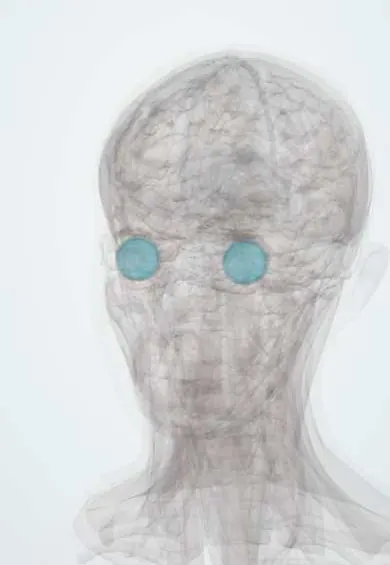
Who gets it
About 1 in 7,500 to 9,000 newborns. It is the most common inherited cause of retinal detachment in children and is often underdiagnosed.
Causes
- COL2A1 (type 1, most common; membranous vitreous; some families have eye-only disease)
- COL11A1 (type 2; beaded vitreous, more hearing loss)
- COL11A2 (no eye involvement; hearing and joint features)
- Recessive forms: COL9A1, COL9A2, COL9A3
Risk factors
- An affected parent (autosomal dominant, 50% risk for each child)
- A newborn with Pierre Robin sequence or cleft palate
Pathophysiology
Abnormal type II and XI collagen weakens the vitreous gel, cartilage and inner ear. The vitreous liquefies early but stays firmly attached at its base, so traction causes large tears at the posterior border of the vitreous base; abnormal cartilage produces the facial, palatal and joint features.
Classification
- Type 1 (COL2A1): membranous vitreous
- Type 2 (COL11A1): beaded vitreous
- Type 3 (COL11A2): non-ocular
- Autosomal recessive types (COL9A1, COL9A2, COL9A3)
Symptoms and signs
- High myopia from early childhood
- Flashes, floaters or a curtain over the vision (retinal detachment)
- Hearing loss (sensorineural or conductive)
- Joint pain, hypermobility and early osteoarthritis
- Feeding or breathing problems at birth (Pierre Robin sequence)
Diagnosis
- Clinical
- Optically empty vitreous with membranes or beaded fibres, radial perivascular lattice degeneration, early wedge-shaped or quadrant cortical cataract, flat midface, small jaw, cleft palate and joint hypermobility.
- Tests
- Cycloplegic refraction; audiometry; genetic testing for COL2A1, COL11A1 and COL11A2.
- Imaging
- Widefield retinal imaging, and B-scan ultrasound when the view is poor; joint X-rays may show epiphyseal dysplasia.
Differential diagnosis
- Wagner syndrome (VCAN; eye-only disease)
- Marfan syndrome
- Isolated high myopia
- Other type II collagen disorders (Kniest dysplasia)
- Knobloch syndrome
Young patient with high myopia and retinal detachment risk: which one?
| Condition | Gene | Vitreous | Retinal detachment | Lens | Rest of the body |
|---|---|---|---|---|---|
| Stickler syndrome | COL2A1, COL11A1, COL11A2 (collagen II and XI) | Optically empty, with membranes or beaded fibres | Common; giant tears | Early wedge or quadrant cortical cataract | Flat midface, cleft palate, deafness, early arthritis |
| Marfan syndrome | FBN1 (fibrillin-1) | Usually normal | More common than average, less than in Stickler | Lens dislocation, usually upward | Tall, long limbs, aortic root dilatation |
| Wagner syndrome | VCAN (versican) | Optically empty, with veils | Less common than in Stickler | Early cataract | None; eye-only disease |
Goal of treatment
Prevent blindness from retinal detachment and manage the craniofacial, hearing and joint problems with a multidisciplinary team.
Treatment
- Procedure
- Prophylactic 360-degree cryotherapy or laser of the peripheral retina in selected patients, especially type 1, to lower the risk of giant tear detachment.
- Surgical
- Scleral buckling or vitrectomy for detachments, which are often complex; cleft palate repair and airway procedures for Pierre Robin sequence.
- Conservative
- Glasses or contact lenses, hearing aids and physiotherapy for the joints.
- Follow-up
- Lifelong dilated retinal exams, teaching patients the warning symptoms of detachment, and genetic counselling.
Complications
- Rhegmatogenous retinal detachment and giant retinal tears
- Cataract
- Glaucoma
- Amblyopia
- Hearing loss
- Early osteoarthritis
Common traps
- High myopia in a young child is only a refractive problem: very high early myopia, especially with cleft palate or a flat face, calls for a vitreous examination for Stickler syndrome
- No systemic features rules out Stickler syndrome: some COL2A1 families have eye-only disease, and the membranous vitreous is the clue
- Stickler detachments behave like ordinary myopic detachments: they often come from giant tears at the posterior border of the vitreous base, can affect both eyes and are surgically complex
- Stickler and Marfan syndromes are separated by the myopia: both cause myopia and detachment, but Marfan brings lens dislocation, tall stature and aortic root disease, while Stickler brings cleft palate, deafness and an optically empty vitreous
- Pierre Robin sequence is only an airway and feeding problem: every infant with it should be checked for Stickler syndrome, because early eye care prevents blindness
Prognosis
The lifetime risk of retinal detachment is high, around half of patients with type 1. Outcomes are better with prophylaxis and early repair; life expectancy is normal.
Prevention
- Screen every infant with Pierre Robin sequence for Stickler syndrome
- Prophylactic retinal treatment and education about detachment symptoms
Test yourself
Think of the answer, then tap the question to see it.
A newborn has a small jaw, cleft palate and noisy breathing. Which syndrome should the eye team look for, and why?
Stickler syndrome, because Pierre Robin sequence is a common way it presents and the child faces a high lifetime risk of retinal detachment.
Why do eyes with Stickler syndrome develop giant retinal tears?
The vitreous liquefies early but stays firmly attached at its base, so traction tears the retina along the posterior border of the vitreous base.
A very myopic child with an optically empty, membranous vitreous has no systemic features. Can this still be Stickler syndrome?
Yes; some COL2A1 families have eye-only disease, and genetic testing confirms it.
Why is prophylactic peripheral retinal treatment offered to selected type 1 patients?
Their lifetime detachment risk is around one in two; 360-degree cryotherapy or laser lowers the risk of giant tear detachment.
How does the lens clue differ between Stickler and Marfan syndromes?
Stickler gives an early wedge-shaped or quadrant cortical cataract; Marfan gives lens dislocation, typically upward and outward.
Connections
Educational information, not medical advice. For any symptom, see a health professional.
Sources: Stickler Syndrome (EyeWiki (American Academy of Ophthalmology), 2025); Stickler syndrome: MedlinePlus Genetics (MedlinePlus, US National Library of Medicine, 2025); Pierre Robin Syndrome (StatPearls Publishing (NCBI Bookshelf), 2023).
Spotted a mistake?We are always improving for the community. Tell us what is wrong and our team will review it.Report a mistake
Representative model, for educational purposes only.