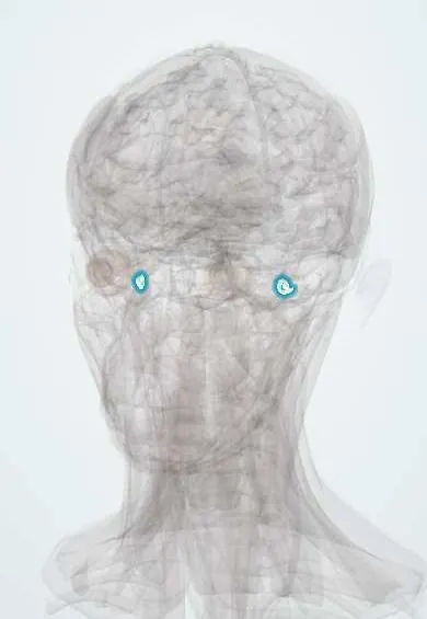
Cochlea !
The cochlea is the snail-shaped, fluid-filled spiral of the inner ear; it turns sound waves into nerve signals, and a cochlear implant sits inside it.
Opens in the Durgical app, no sign-up needed.
What it is
A snail-shaped spiral of fluid-filled tubes in the inner ear.
What it does
Turns sound waves into nerve signals; a cochlear implant sits inside it.
Key facts
- Where
- Inner ear, petrous temporal bone; 2.5 turns
- Blood supply
- Labyrinthine artery (AICA/basilar)
- Nerve
- Cochlear part of VIII
- Function
- Hearing (organ of Corti); base = high pitch, apex = low
Relations
- Contents
- Bony spiral around a central axis (modiolus) that holds the spiral ganglion; scala vestibuli, cochlear duct with the organ of Corti on the basilar membrane, and scala tympani
- Lateral
- Middle ear: the basal turn bulges as the promontory; the oval window (stapes footplate) above it opens to the vestibule and the round window closes the scala tympani
- Behind
- Vestibule and semicircular canals, the rest of the bony labyrinth
- Medial
- Fundus of the internal acoustic meatus, where cochlear nerve fibres leave through its anteroinferior part
- In front
- Carotid canal with the internal carotid artery, close by in the petrous bone
How to find it
Deep in the petrous part of the temporal bone and cannot be examined directly. It is reached through the middle ear: the promontory and round window niche are seen once the eardrum is lifted, and a cochlear implant electrode is passed through the round window into the scala tympani.
Examination
- Rinne positive (air louder than bone) in both ears and Weber lateralising to the better ear: sensorineural loss
- Pure-tone audiometry: air and bone conduction thresholds drop together with no air-bone gap
- Otoacoustic emissions: absent when outer hair cells are damaged; used to screen newborns
On imaging
CT of the temporal bone shows the bony cochlea (two and a half turns), malformations and the lucent ring of cochlear otosclerosis; high-resolution T2-weighted MRI shows the fluid inside it and the cochlear nerve, and confirms the cochlea is open before an implant.
Common problems
- Age-related and noise-induced hearing loss: the sensory hair cells wear out.
- Sudden sensorineural hearing loss: an emergency.
- Tinnitus (ringing).
How it is diagnosed
Audiometry; MRI if one side is affected.
Treatment
- Hearing protection; hearing aids.
- Urgent steroids for sudden hearing loss.
- Cochlear implant for severe to profound loss.
Clinical notes
- Cochlear implant: an electrode array in the scala tympani stimulates the spiral ganglion directly, bypassing lost hair cells; it needs an intact cochlear nerve
- After bacterial meningitis the cochlea can fill with bone (labyrinthitis ossificans); hearing is checked early so an implant can be placed before it closes
- Ototoxic drugs such as aminoglycosides and cisplatin damage outer hair cells, high frequencies first
- Ménière disease: excess endolymph (hydrops) with episodic vertigo, fluctuating low-frequency hearing loss, tinnitus and aural fullness
Development
The membranous labyrinth grows from the otic placode (ectoderm) via the otic vesicle; the surrounding otic capsule ossifies into the bony labyrinth, adult-sized at birth.
Memory aid
Endolymph is like the inside of a cell (high potassium); perilymph is like plasma or CSF (high sodium).
Related conditions
- Ménière disease
- Presbycusis (age-related hearing loss)
- Hearing loss (conductive and sensorineural, including sudden hearing loss)
- Otosclerosis
Test yourself
Think of the answer, then tap the question to see it.
What are the three spaces of the cochlea and their fluids?
Scala vestibuli and scala tympani hold perilymph and meet at the helicotrema; the cochlear duct between them holds potassium-rich endolymph and the organ of Corti.
What do otoacoustic emissions test, and where are they used?
Outer hair cell function in the cochlea; they are the basis of newborn hearing screening.
What audiogram patterns are typical of noise-induced hearing loss and presbycusis?
Noise: a notch near 4 kHz; presbycusis: bilateral, symmetric high-frequency loss starting at the base of the cochlea.
Why does unilateral or asymmetric sensorineural hearing loss need imaging?
To exclude a vestibular schwannoma.
Where is a cochlear implant electrode placed, and what does it require?
In the scala tympani, via the round window; it stimulates the spiral ganglion directly and needs an intact cochlear nerve.
Educational information, not medical advice. For any symptom, see a health professional.
Its names
- Official Latin (TA2)
- cochlea TA2 6964
- Official Spanish (DeCS)
- Cóclea DeCS D003051
- English (TA2)
- Cochlea
Related
Sources: Terminologia Anatomica (TA2); DeCS; Inner Ear Anatomy and Cochlear Physiology; Neuroanatomy, Cranial Nerve 8 (Vestibulocochlear).
Spotted a mistake?We are always improving for the community. Tell us what is wrong and our team will review it.Report a mistake
Representative model, for educational purposes only.