Conditions · Metabolism (biochemistry)
Tay–Sachs disease !
In Tay–Sachs disease a lipid called GM2 builds up in neurons; the baby stops progressing, loses skills and loses sight.
Updated 2026-11-01
Structures involved
Tap one to see what it is and open it in 3D.
In biochemistry
- Lysosomal ganglioside breakdown: Step 2 · β-Hexosaminidase A (HEXA α + HEXB β subunits) with the GM2 activator
- Sphingolipids and sphingolipidoses
- Lysosomal storage diseases
- GM2 ganglioside
- Hexosaminidase A (β-hexosaminidase A)
- Hexosaminidase B (β-hexosaminidase B)
In histology
Why it happens
- How a single gene causes disease: enzymes, receptors and structural proteins
- Intracellular accumulations: cholesterol, proteins, hyaline, glycogen
What it is
An autosomal recessive GM2 gangliosidosis caused by deficiency of the α subunit of hexosaminidase A (HEXA gene). It affects almost only the nervous system.
Who gets it
Very rare in the general population. The variants are more common in Ashkenazi Jews, French-Canadian communities of Quebec, the Amish and the Cajun people of Louisiana; carrier screening has greatly reduced cases in these groups.
Causes
- Biallelic variants of the HEXA gene (autosomal recessive)
Risk factors
- Carrier parents
- Belonging to a population with a high carrier frequency
Pathophysiology
Without hexosaminidase A, GM2 ganglioside from neuronal membranes is not converted to GM3 and fills neuronal lysosomes (membranous cytoplasmic bodies). Neurons swell, lose function and die. In the retina, lipid-laden ganglion cells look whitish around the fovea, which lacks them and stands out red (cherry-red spot). There is no major visceral storage, so the liver and spleen do not enlarge.
Classification
- Infantile (classic): onset at 3–6 months, the most common
- Juvenile: onset in childhood, slower course
- Late-onset or adult: weakness, ataxia and psychiatric symptoms
Symptoms and signs
- Exaggerated startle to noise
- Loss of motor skills already gained (head control, sitting)
- Hypotonia that later turns into spasticity
- Progressive loss of vision
- Seizures
- Enlarging head (macrocephaly)
Diagnosis
- Clinical
- An infant with developmental regression, exaggerated startle and a cherry-red spot on fundoscopy, without hepatosplenomegaly.
- Tests
- Low hexosaminidase A activity with normal or high total hexosaminidase in serum or leukocytes; HEXA gene testing; electroencephalogram if there are seizures.
- Imaging
- Brain MRI with white matter and thalamic changes and, later, atrophy; CT with hyperdense thalami.
Treatment
- Acute
- Management of seizures and aspiration pneumonia.
- Medical
- No treatment stops the disease; anticonvulsants and control of drooling and spasticity are used. Gene therapy is under investigation.
- Rehabilitation
- Physiotherapy, swallowing therapy and nutritional support (sometimes a gastrostomy tube).
- Conservative
- Palliative care and family support, with genetic counselling.
Complications
- Aspiration pneumonia
- Status epilepticus
- Blindness
- Malnutrition
Prognosis
The infantile form usually leads to death between ages 2 and 5. Juvenile and adult forms progress more slowly.
Prevention
- Carrier screening in at-risk populations
- Genetic counselling and prenatal diagnosis when both parents are carriers
Test yourself
Think of the answer, then tap the question to see it.
Which enzyme is missing in Tay–Sachs disease?
Hexosaminidase A, from variants in the HEXA gene (α subunit).
Why does Tay–Sachs not cause hepatosplenomegaly, unlike Niemann–Pick?
GM2 turns over mainly in neurons; there is no major visceral storage.
Which fundoscopic finding is typical of the infantile form?
A cherry-red spot at the macula.
Which early sign stands out in an infant with Tay–Sachs?
An exaggerated startle to noise, before motor regression.
Related conditions
Educational information, not medical advice. For any symptom, see a health professional.
Sources: Tay-Sachs disease: MedlinePlus Genetics (MedlinePlus, US National Library of Medicine, 2026); HEXA gene: MedlinePlus Genetics (MedlinePlus, US National Library of Medicine, 2026); Textbook of Biochemistry for Medical Students, 6th ed. (Jaypee Brothers Medical Publishers (Vasudevan DM, Sreekumari S, Vaidyanathan K), 2011).
Spotted a mistake?We are always improving for the community. Tell us what is wrong and our team will review it.Report a mistake
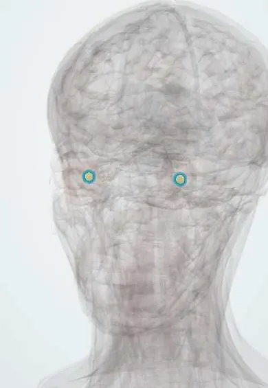
Representative model, for educational purposes only.