
Traumatic optic neuropathy
A blow to the head or brow can damage the optic nerve even when the eye looks perfect. Vision drops and the pupil reacts poorly to light.
What it is
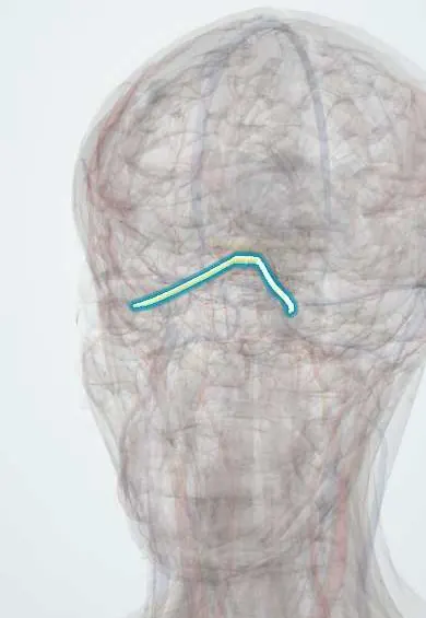
Loss of vision from injury to the optic nerve after head, brow or orbital trauma. Most cases are indirect: the force of the blow is transmitted to the nerve where it is tethered in the bony optic canal, which bruises or shears it without any visible wound. Direct injury from a fracture fragment, a penetrating object or a haematoma is less common. The eye itself often looks normal.
Who gets it
Occurs in a small percentage of closed head injuries, mostly in young men after road traffic accidents, falls and assaults; often in patients who are unconscious or have other injuries, so it is easily missed.
Causes
- Indirect injury: frontal or brow impact transmitting force to the optic canal (most common)
- Direct injury: fracture fragment in the canal, penetrating injury
- Compression by orbital or optic nerve sheath haemorrhage
- Avulsion of the nerve from the globe in severe rotational trauma
Risk factors
- Road traffic accidents, especially motorcycle and bicycle
- Falls and assaults with brow impact
- Severe head injury and loss of consciousness
Pathophysiology
At impact, axons in the canal are sheared and small vessels torn. A second phase follows: swelling in the tight canal compresses the nerve and its blood supply, and ischaemia and inflammation kill more ganglion cell axons. Lost axons do not regrow, so the disc becomes pale (optic atrophy) about four to six weeks later.
Classification
- Indirect (concussive force to the canal) or direct (fragment, penetration, avulsion)
- Anterior (near the globe: early disc swelling or haemorrhage) or posterior (behind the globe: normal disc at first)
Symptoms and signs
- Reduced vision in one eye after head or brow trauma, often immediately
- Dim or washed-out colours
- Field loss
- Sometimes no complaint in an unconscious or confused patient
Diagnosis
- Clinical
- A clinical diagnosis: visual loss with a relative afferent pupillary defect after trauma, in an eye with no other cause. The disc is usually normal at first in posterior injury and turns pale in four to six weeks. In the unconscious patient the swinging flashlight test is the key sign.
- Tests
- Visual acuity, colour vision and visual fields when the patient can cooperate; visual evoked potentials in selected unconscious patients.
- Imaging
- Thin-cut CT of the orbits and optic canal to look for a canal fracture, a bone fragment or an orbital or sheath haemorrhage; MRI is rarely needed acutely.
Differential diagnosis
- Retrobulbar haemorrhage with orbital compartment syndrome
- Globe rupture, vitreous haemorrhage or retinal detachment
- Commotio retinae
- Chiasmal or cortical injury (field loss without a unilateral pupil defect)
- Functional visual loss (normal pupils)
Visual loss after trauma: where is the damage?
| Clue | Traumatic optic neuropathy | Retrobulbar haemorrhage | Globe injury | Cortical or chiasmal injury |
|---|---|---|---|---|
| Eye on inspection | Looks normal | Tense, bulging, will not move | Hyphema, misshapen pupil, red reflex lost | Looks normal |
| Afferent pupil defect | Yes, one side | Yes | Only with severe damage | No (both sides affected equally) |
| Fundus | Normal disc at first, pale weeks later | May be normal; artery may pulse | Bleeding, detachment or no view | Normal |
| Urgency | Assess, image, discuss; no proven treatment | Minutes: canthotomy | Same day: protect the globe | Neurology and neurosurgery care |
Goal of treatment
Rule out and treat compressive causes, then support recovery without causing harm.
Treatment
- Acute
- Exclude retrobulbar haemorrhage first: a tense orbit with falling vision needs lateral canthotomy at once.
- Follow-up
- Observation is the standard option: a substantial share of patients improve on their own, and no treatment has been shown to beat it.
- Medical
- High-dose corticosteroids are not recommended routinely: they have not shown benefit for the nerve and increased mortality in head injury.
- Surgical
- Optic canal decompression is considered only in selected cases, such as a bone fragment impinging on the nerve or delayed worsening, after specialist discussion.
Complications
- Permanent visual loss and optic atrophy
- Bilateral blindness in severe injuries
- Missed associated brain or orbital injuries
Common traps
- A normal-looking eye and disc rule out an optic nerve injury: in posterior injury the disc stays normal for weeks; the afferent pupil defect is the early sign.
- High-dose steroids are the standard treatment for a traumatic optic nerve injury: they have no proven benefit, and in head injury they increased mortality.
- Every traumatic optic neuropathy should go for decompression surgery: neither surgery nor steroids has beaten observation; surgery is reserved for selected compressive cases.
- If the pupil defect is present, the cause must be the optic nerve itself: a retrobulbar haemorrhage gives the same defect and is treatable within minutes, so check the orbit first.
- An unconscious patient cannot be assessed for vision: the swinging flashlight test needs no cooperation and should be part of the trauma survey.
Prognosis
Variable: some patients recover useful vision on their own; no light perception at presentation, a canal fracture and no improvement in the first days predict a poor outcome.
Prevention
- Helmets for motorcyclists and cyclists
- Seat belts and airbags
- Fall prevention
Test yourself
Think of the answer, then tap the question to see it.
After a motorcycle crash with a blow to the right brow: right vision 20/400, a normal eye and disc, a right afferent pupil defect. Likely diagnosis?
Traumatic optic neuropathy, an indirect injury at the optic canal; the disc looks normal early on.
Why is the optic nerve injured at the optic canal rather than inside the orbit?
In the canal it is fixed to the bone, so the transmitted force shears it and swelling has no room; in the orbit the nerve is slack and mobile.
Why are high-dose corticosteroids not given routinely?
They have no proven benefit over observation, and in head injury they were linked to higher mortality.
Before labelling visual loss after trauma as traumatic optic neuropathy, which treatable condition must be excluded?
Retrobulbar haemorrhage with orbital compartment syndrome, treated by immediate lateral canthotomy.
Connections
Educational information, not medical advice. For any symptom, see a health professional.
Sources: Traumatic Optic Neuropathy (EyeWiki, American Academy of Ophthalmology, 2025); Retrobulbar Hematoma (StatPearls Publishing (NCBI Bookshelf), 2022); Optic Atrophy (StatPearls Publishing (NCBI Bookshelf), 2024).
Spotted a mistake?We are always improving for the community. Tell us what is wrong and our team will review it.Report a mistake
Representative model, for educational purposes only.