
Periorbital haematoma (black eye)
A black eye is usually just a bruise, but it can hide an injured eyeball, an orbital fracture or, in both eyes, a fracture of the skull base.
What it is
Bleeding into the loose skin of the eyelids and around the orbit, most often from a direct blow. The bruise itself is harmless and fades over one to two weeks. What matters is what lies under it: an injured globe, a fracture of the orbital walls, bleeding behind the eye, or, when both eyes are bruised without a blow to the face, a fracture of the anterior skull base.
Who gets it
Very common after assaults, falls, sports and road traffic injuries; bilateral bruising after head injury is a classic sign of anterior skull base fracture.
Causes
- Direct blunt trauma to the eye or brow
- Nasal or orbital fracture
- Anterior cranial fossa (basilar skull) fracture, with blood tracking into both orbits
- Facial or eyelid surgery
- Less often without trauma: anticoagulants or bleeding disorders, amyloidosis, metastatic neuroblastoma in children
Risk factors
- Anticoagulant or antiplatelet treatment
- Contact sports, assaults and falls in older adults
- Bleeding disorders
Pathophysiology
Blunt force tears small vessels in the lid and orbit. The eyelid skin has almost no subcutaneous fat and a loose plane under it, so blood spreads easily and gravity pulls it downwards, sometimes into the other lid across the nose. Blood from an anterior skull base fracture tracks into both orbits but stays within the orbital septum, which gives the ringed 'raccoon' appearance after a delay.
Classification
- Unilateral after a local blow: lid bruise with or without globe or orbital injury
- Bilateral after head injury (raccoon eyes): suspect anterior skull base fracture
- With tense proptosis and visual loss: retrobulbar haemorrhage (orbital compartment syndrome)
- Spontaneous, without trauma: look for a bleeding disorder or tumour
Symptoms and signs
- Swollen, blue-purple eyelids that may close the eye
- Pain and tenderness
- Blurred or double vision when the globe or an eye muscle is injured
- Numb cheek and upper lip when the infraorbital nerve is injured
Diagnosis
- Clinical
- Always open the lids and examine the eye itself: visual acuity, pupils (relative afferent defect), eye movements, globe position, anterior chamber, and sensation over the cheek. Gently, without pressing on the globe, if an open globe is possible. Look for raccoon eyes, bruising behind the ear and fluid from the nose or ear.
- Tests
- Clear nasal or ear fluid can be checked for beta-2 transferrin (cerebrospinal fluid); coagulation tests if bruising is spontaneous or excessive.
- Imaging
- CT of the orbits and face (thin cuts) for suspected fracture, retrobulbar haemorrhage or a foreign body; CT of the head when a skull base fracture or brain injury is possible. Imaging must not delay decompression of a tense orbit with falling vision.
Differential diagnosis
- Orbital or preseptal cellulitis (fever, no trauma)
- Allergic or angio-oedema of the lids (itchy, no bruising)
- Subconjunctival haemorrhage alone
- Spontaneous orbital bleeding from a vascular lesion or tumour
One black eye or two: what lies under the bruise
| Clue | Simple lid bruise | Orbital floor fracture | Anterior skull base fracture | Retrobulbar haemorrhage |
|---|---|---|---|---|
| Onset | Immediate after a blow | Immediate after a blow | Often delayed one to three days after head injury | Minutes to hours after the injury |
| Eyes affected | One | One | Usually both (raccoon eyes) | One |
| Vision and pupil | Normal | Usually normal | Usually normal | Falling vision, afferent pupillary defect |
| Key extra signs | None | Upgaze diplopia, numb cheek, sunken eye | Clear fluid from the nose, loss of smell, bruising behind the ear | Tense bulging eye that will not move, high pressure |
| What to do | Cold compresses, reassurance | CT orbits, ophthalmology, no nose blowing | CT head, neurosurgery | Lateral canthotomy within the hour |
Goal of treatment
Find and treat what lies under the bruise; the bruise itself only needs comfort measures.
Treatment
- Conservative
- Cold compresses in the first two days, head raised, simple pain relief; avoid antiplatelet painkillers when bleeding is a concern.
- Acute
- Lateral canthotomy and cantholysis at once for a tense orbit with falling vision; it is done before imaging.
- Medical
- With an orbital fracture: no nose blowing, and antibiotics or decongestants as decided by the treating team.
- Follow-up
- Ophthalmology review for any change in vision, pupils or eye movement; neurosurgical care for a skull base fracture.
Complications
- Missed globe rupture or hyphema
- Orbital compartment syndrome and blindness
- Persistent double vision or enophthalmos from an untreated fracture
- Cerebrospinal fluid leak and meningitis after skull base fracture
Common traps
- A black eye only needs ice: the swollen lids hide the eye, so measure vision and check the pupils and movements in every case.
- Raccoon eyes come from a blow to both eyes: they usually appear a day or more after a head injury with no facial blow, because blood tracks down from a skull base fracture.
- The bulging eye needs a CT before anything else: a tense orbit with falling vision is decompressed by lateral canthotomy first, because irreversible vision loss can begin after roughly 90–120 minutes of ischaemia.
- Forcing the swollen lids open is part of a thorough exam: if the globe may be open, pressure can push its contents out; open the lids gently with retractors or leave it to ophthalmology.
- A child's black eye after a fall is always accidental: bruising that does not fit the story, or both eyes in a young child, needs a safeguarding assessment.
Prognosis
The bruise fades in one to two weeks with yellow-green colour changes; the outcome depends on any globe, orbital or brain injury beneath it.
Prevention
- Protective eyewear in sport and work
- Seat belts and airbags
- Fall prevention in older adults on anticoagulants
Test yourself
Think of the answer, then tap the question to see it.
Two days after falling and hitting the back of his head, a man has bruising around both eyes and clear fluid from his nose. What is the likely injury?
An anterior skull base (basilar) fracture with a cerebrospinal fluid leak; he needs CT of the head and neurosurgical care.
Why must you open the swollen lids of every black eye?
Because the bruise can hide a globe rupture, hyphema or retrobulbar haemorrhage, which change management and threaten sight.
After a punch, a patient cannot look up and the cheek is numb. Which structure is broken and which nerve is affected?
The orbital floor, with the infraorbital nerve that runs in it.
A black eye becomes tense and bulging, vision falls and the pupil shows an afferent defect. What do you do first?
Lateral canthotomy and cantholysis at once, before imaging, to decompress the orbit.
Connections
Educational information, not medical advice. For any symptom, see a health professional.
Sources: Traumatic Orbital Compartment Syndrome in Children: Systematic Review and Descriptive Summary (Children (Basel) (Europe PMC open access), 2026); Basilar Skull Fractures (StatPearls Publishing (NCBI Bookshelf), 2023); Orbital Floor Fracture (StatPearls Publishing (NCBI Bookshelf), 2023); Retrobulbar Hematoma (StatPearls Publishing (NCBI Bookshelf), 2022); Lateral Orbital Canthotomy (StatPearls Publishing (NCBI Bookshelf), 2023).
Spotted a mistake?We are always improving for the community. Tell us what is wrong and our team will review it.Report a mistake
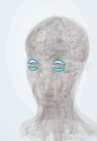
Representative model, for educational purposes only.