Conditions · Infectious and tropical diseases
Viral haemorrhagic fevers (Ebola and others) !
Viral haemorrhagic fevers are severe viral illnesses such as Ebola that cause fever, organ failure and sometimes bleeding, and need strict isolation.
Updated 2026-11-01
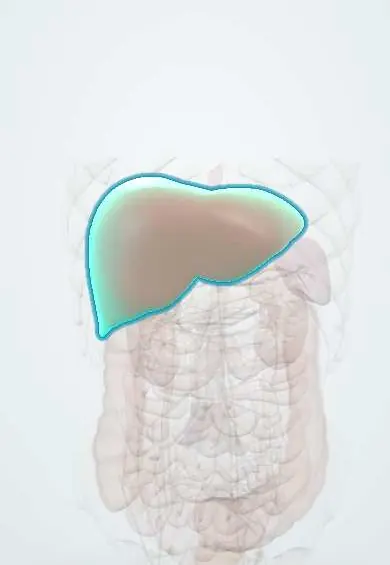
Structures involved
Tap one to see what it is and open it in 3D.
Microorganisms that cause it
- Ebola and Marburg viruses (filoviruses)
- Hantavirus
- Arenaviruses (Lassa and the South American haemorrhagic fevers)
Why it happens
- Disseminated intravascular coagulation: how consumption causes both clots and bleeding
- Shock: types, stages and the organs it damages
- The coagulation cascade
- Functions of the liver
What it is
A group of severe systemic illnesses caused by RNA viruses from several families (filoviruses such as Ebola and Marburg; arenaviruses such as Lassa and the South American viruses; bunyaviruses; flaviviruses such as dengue and yellow fever) that damage blood vessels and can cause shock, organ failure and bleeding.
Who gets it
Ebola and Marburg occur in outbreaks in sub-Saharan Africa, with average case fatality around 50%. South American arenaviruses cause Argentine (Junín), Bolivian (Machupo, Chapare), Venezuelan (Guanarito) and Brazilian (Sabiá) haemorrhagic fevers, linked to wild rodents in rural areas. In Colombia the haemorrhagic fevers seen in practice are mostly dengue and yellow fever; filovirus disease is a risk only in travellers or health workers returning from outbreak zones.
Causes
- Filoviruses: Ebola (Zaire, Sudan, Bundibugyo) and Marburg; reservoir in fruit bats
- South American arenaviruses: Junín, Machupo, Guanarito, Sabiá and Chapare; reservoir in wild rodents (Lassa virus in West Africa)
- Bunyaviruses (Crimean-Congo haemorrhagic fever, hantaviruses) and flaviviruses (dengue, yellow fever)
- Human-to-human spread of filoviruses (and some arenaviruses such as Machupo and Chapare) through blood and body fluids
Risk factors
- Caring for patients or preparing bodies without protective equipment
- Unsafe burial practices with direct contact with the body
- Entering caves or mines with bats; handling bushmeat
- Rural farm work with exposure to rodent urine and droppings (arenaviruses)
- Travel to an area with an active outbreak
Pathophysiology
The viruses infect macrophages and dendritic cells, then endothelium, liver and adrenal glands. Uncontrolled release of cytokines and impaired immune responses increase vascular permeability, causing fluid loss and shock. Liver injury, reduced clotting factor production, thrombocytopenia and disseminated intravascular coagulation explain the bleeding, which is a late and inconstant sign; multi-organ failure is the usual cause of death.
Classification
- Filovirus diseases: Ebola disease and Marburg virus disease
- Arenavirus haemorrhagic fevers: South American (Argentine, Bolivian, Venezuelan, Brazilian, Chapare) and Lassa fever
- Bunyavirus diseases: Crimean-Congo haemorrhagic fever, haemorrhagic fever with renal syndrome, hantavirus pulmonary syndrome
- Flavivirus diseases: severe dengue, yellow fever
Symptoms and signs
- Sudden fever, intense fatigue, myalgia, headache and sore throat
- Vomiting, profuse diarrhoea and abdominal pain (the ‘wet’ phase of Ebola, causing severe fluid loss)
- Rash and conjunctival injection
- Bleeding from gums, nose, puncture sites or the gut, and petechiae, usually late
- Tremor, confusion or seizures in arenavirus and late filovirus disease
Diagnosis
- Clinical
- Suspected from fever plus an epidemiological link (travel, contact with a case, bats, rodents). The patient is isolated immediately and public health is notified before samples are sent; early symptoms resemble malaria, typhoid and dengue, which must also be tested.
- Tests
- RT-PCR of blood in reference laboratories with high biosafety containment; antigen and antibody tests. Laboratory clues: leukopenia then neutrophilia, thrombocytopenia, raised transaminases (AST higher than ALT), kidney injury, electrolyte disturbances and coagulation abnormalities.
- Imaging
- Not diagnostic; bedside ultrasound to assess volume status and organ complications, limited to reduce staff exposure.
Differential diagnosis
- Malaria
- Severe dengue and yellow fever
- Typhoid fever
- Leptospirosis and rickettsial infections
- Bacterial sepsis with disseminated intravascular coagulation
Goal of treatment
Isolate the patient safely, restore and maintain circulation, correct electrolytes and organ failure, use virus-specific therapy where it exists, and stop transmission to staff and contacts.
Treatment
- Follow-up
- Immediate isolation in a single room with dedicated equipment; trained staff in full personal protective equipment (gloves, impermeable gown, face shield or goggles, respirator for aerosol-generating procedures) with supervised donning and removal; strict fluid balance, vital signs and waste handling.
- Acute
- Optimised supportive care: intravenous fluids and electrolyte replacement, management of shock with vasopressors, oxygen, blood products for bleeding, and kidney replacement when needed; this alone improves survival.
- Medical
- Ebola (Zaire ebolavirus): monoclonal antibody therapy (ansuvimab or a three-antibody combination); no approved specific treatment for Sudan, Bundibugyo or Marburg viruses.
- Medical
- Arenaviruses: ribavirin for Lassa fever and considered for South American haemorrhagic fevers; early immune (convalescent) plasma reduces mortality in Argentine haemorrhagic fever.
- Follow-up
- Contact tracing and daily follow-up of contacts for 21 days; safe and dignified burials; Ebola vaccination of contacts and health workers during outbreaks (ring vaccination).
Drugs for this condition
Complications
- Hypovolaemic and distributive shock
- Acute kidney injury and electrolyte disorders (hypokalaemia)
- Liver failure and disseminated intravascular coagulation
- Encephalitis and seizures
- Miscarriage and very high maternal and fetal mortality
- Long-term sequelae in survivors (uveitis, joint pain, fatigue) and viral persistence in semen and the eye
Prognosis
Average case fatality for Ebola and Marburg is around 50% (range about 25–90% across outbreaks), lower with early intensive supportive care and, for Ebola Zaire, monoclonal antibodies. Argentine haemorrhagic fever has a fatality of 15–30% untreated, much lower with early immune plasma.
Prevention
- Standard, contact and droplet precautions and full protective equipment for every suspected case
- Ebola vaccination for outbreak response (ring vaccination of contacts and front-line workers); a live attenuated vaccine against Junín virus is used in endemic Argentina
- Rodent control and sealing homes and food stores in arenavirus areas
- Avoid contact with bats, bushmeat and bodies of the deceased; safe burials
- Screening of travellers from outbreak areas and 21-day follow-up of exposed people
Test yourself
Think of the answer, then tap the question to see it.
What is the incubation period of Ebola, and why does it matter for contact follow-up?
2 to 21 days, so contacts are followed daily for 21 days after their last exposure.
Can a person with Ebola transmit the virus before symptoms appear?
No; transmission starts with symptoms, through blood and body fluids, and is highest in late illness and after death.
Which isolation precautions does a nurse use for a patient with suspected Ebola?
A single room with dedicated equipment, standard, contact and droplet precautions with full PPE, respirator for aerosols, and supervised donning and doffing.
Which virus family causes the South American haemorrhagic fevers, and what is the reservoir?
Arenaviruses (Junín, Machupo, Guanarito, Sabiá, Chapare), carried by wild rodents.
Which treatment reduces mortality in Argentine haemorrhagic fever?
Early immune (convalescent) plasma, ideally in the first week of illness.
Related conditions
- Dengue
- Yellow fever
- Malaria
- Leptospirosis
- Typhoid (enteric) fever
- Hantavirus pulmonary syndrome
- Disseminated intravascular coagulation
- Sepsis and septic shock
- Acute kidney injury
- Acute liver failure
- Rickettsial infections (spotted fever)
Educational information, not medical advice. For any symptom, see a health professional.
Sources: Ebola disease (World Health Organization, 2025); Marburg virus disease (World Health Organization, 2025); About Viral Hemorrhagic Fevers | Viral Hemorrhagic Fevers (VHFs) | CDC (Centers for Disease Control and Prevention, 2024).
Spotted a mistake?We are always improving for the community. Tell us what is wrong and our team will review it.Report a mistake
Representative model, for educational purposes only.