
Von Hippel-Lindau disease
An inherited condition with blood-vessel tumours in the eye and brain and a high risk of kidney cancer. Lifelong check-ups save sight and lives.
What it is
An autosomal dominant cancer syndrome from mutation of the VHL tumour suppressor gene (chromosome 3p). Retinal haemangioblastomas are often the first sign in young people; cerebellar and spinal haemangioblastomas, clear cell renal carcinoma, phaeochromocytoma and pancreatic and endolymphatic sac tumours follow.
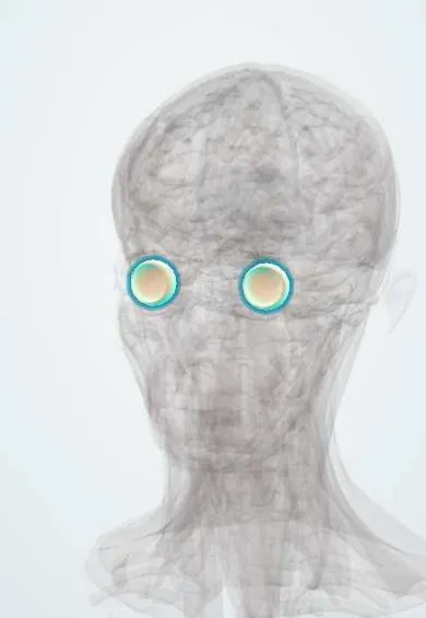
See it in 3D
Who gets it
About 1 in 36 000 people. Penetrance is high; about one in five cases are new mutations without family history. Retinal tumours affect most patients and are bilateral in about half.
Causes
- Germline mutation of the VHL gene (chromosome 3p25), with a second hit in the tissue
- Autosomal dominant inheritance
Risk factors
- Affected parent (50% risk to each child)
Pathophysiology
Loss of VHL protein stops the breakdown of hypoxia-inducible factor, so cells act as if starved of oxygen and overproduce VEGF and erythropoietin. This drives highly vascular tumours. In the retina, a red haemangioblastoma with a dilated feeding artery and draining vein leaks exudate and fluid and can cause traction.
Classification
- Retinal haemangioblastoma: peripheral (endophytic, with feeder vessels) or juxtapapillary (at the optic disc, hard to treat)
- VHL type 1: low phaeochromocytoma risk; type 2: phaeochromocytoma risk (2A, 2B with renal cancer, 2C phaeochromocytoma only)
Symptoms and signs
- Often none when tumours are small
- Blurred vision from macular exudate or fluid
- Floaters or field loss from detachment or vitreous haemorrhage
- Systemic: headache, unsteadiness, blood in the urine, episodes of palpitations and sweating, hearing loss
Diagnosis
- Clinical
- Dilated fundus examination of the whole retina, including the far periphery; a family history and one typical tumour, or two tumours without family history, make the diagnosis.
- Tests
- Genetic testing for the VHL mutation in the patient and at-risk relatives; plasma or urine metanephrines for phaeochromocytoma.
- Imaging
- Fluorescein angiography (rapid filling and leakage, shows small tumours); OCT for macular fluid; MRI of the brain and spine and of the abdomen for systemic tumours.
Differential diagnosis
- Coats disease
- Retinal vasoproliferative tumour
- Retinal cavernous haemangioma
- Racemose haemangioma (Wyburn-Mason)
- Retinal macroaneurysm
Red retinal lesion with abnormal vessels: which one?
| Condition | Lesion | Vessels | Typical patient | Systemic link |
|---|---|---|---|---|
| Von Hippel-Lindau disease | Round red-orange tumour, peripheral or at the disc | Paired dilated feeding artery and draining vein | Teenager or young adult, often bilateral, family history | Kidney, cerebellum, spine, adrenal, pancreas, inner ear |
| Coats disease | No discrete tumour; massive yellow exudate | Irregular telangiectatic, aneurysmal vessels | Young boy, one eye | None |
| Retinal vasoproliferative tumour | Yellow-pink peripheral mass, usually inferior | Only mildly dilated feeders | Middle-aged adult, often after other eye disease | None |
| Racemose haemangioma (Wyburn-Mason) | No mass; tangle of tortuous vessels | Direct artery-to-vein connections without capillaries | Young person, one eye | Similar malformation in the brain on the same side |
Goal of treatment
Find and treat retinal tumours while small, and detect systemic tumours early through lifelong surveillance.
Treatment
- Procedure
- Laser photocoagulation for small peripheral tumours; cryotherapy for larger or anterior ones; photodynamic therapy or radiotherapy for selected juxtapapillary tumours.
- Surgical
- Vitrectomy for tractional detachment or non-clearing vitreous haemorrhage.
- Medical
- Belzutifan, an oral HIF-2-alpha inhibitor, can shrink VHL-related kidney, brain, pancreatic and retinal tumours under specialist care.
- Follow-up
- Yearly eye examination from infancy, with regular blood pressure, metanephrine, abdominal MRI, brain and spine MRI and hearing checks per surveillance protocols.
Complications
- Exudative or tractional retinal detachment
- Vitreous haemorrhage
- Neovascular glaucoma and loss of the eye
- Renal cell carcinoma (main cause of death)
- Cerebellar haemangioblastoma
Common traps
- A single retinal haemangioblastoma in a young adult is a local eye problem: it is often the first sign of VHL disease, so genetic testing and systemic screening follow
- The eye tumours are what kill patients with VHL: clear cell renal carcinoma is the main cause of death, with central nervous system haemangioblastomas next
- Phaeochromocytoma screening can wait for symptoms: an unsuspected tumour can cause a hypertensive crisis under anaesthesia, so metanephrines are checked before any surgery
- Examining the posterior pole is enough: small peripheral tumours are the easiest to treat, so the far periphery is examined, with fluorescein angiography for tiny lesions
Prognosis
Structured surveillance has improved survival markedly; renal cancer and central nervous system tumours drive mortality. Most eyes keep useful vision if tumours are treated early.
Prevention
- Genetic testing of relatives and cascade screening
- Lifelong surveillance from early childhood
Test yourself
Think of the answer, then tap the question to see it.
A 19-year-old has a red peripheral retinal tumour with a dilated artery and vein. Which tests follow?
VHL gene testing, plasma or urine metanephrines, MRI of the brain, spine and abdomen, and fluorescein angiography of both eyes.
Why are VHL tumours so rich in blood vessels?
Without VHL protein, HIF accumulates and switches on VEGF, as if the tissue were starved of oxygen.
A patient with VHL is booked for cerebellar surgery. What must be checked first, and why?
Metanephrines, because a hidden phaeochromocytoma can cause a hypertensive crisis under anaesthesia.
Why are small peripheral retinal tumours treated rather than watched?
They tend to grow and leak; small ones respond well to laser, while large or juxtapapillary ones are hard to treat.
Why does a patient with VHL need surveillance for life even after every tumour is treated?
Every cell carries the germline mutation, so new tumours keep arising in the eye and elsewhere.
Why it happens
- Cystic pancreatic neoplasms
- Non-neoplastic pancreatic cysts: congenital cysts and pseudocysts
- Familial tumour syndromes of the nervous system
- Clear cell renal cell carcinoma
In dermatology
In neurology
Connections
Educational information, not medical advice. For any symptom, see a health professional.
Sources: Retinal Vascular Anomalies (VHL, Cavernous Hemangioma, Wyburn-Mason) (StatPearls Publishing (NCBI Bookshelf), 2024); Hemangioblastoma (StatPearls Publishing (NCBI Bookshelf), 2025); Von Hippel-Lindau Syndrome (EyeWiki (American Academy of Ophthalmology), 2025).
Spotted a mistake?We are always improving for the community. Tell us what is wrong and our team will review it.Report a mistake
Representative model, for educational purposes only.