Ischaemic stroke !
An ischaemic stroke happens when a blocked artery cuts off blood to part of the brain; it causes sudden weakness, numbness or trouble speaking.
Updated 2026-10-29
Structures involved
Tap one to see what it is and open it in 3D.
- Middle cerebral artery
- Anterior cerebral artery
- Posterior cerebral artery
- Internal carotid artery
- Circle of Willis
- Basilar artery
- Vertebral artery
- Carotid bifurcation
- Motor cortex
- Broca's area
In biochemistry
- Glutamate (glutamic acid)
- Homocysteine
- LDL (low-density lipoprotein)
- Thromboxane A₂ (TXA₂)
- HMG-CoA reductase
In histology
- Cell injury, apoptosis and necrosis
- Stem cells and tissue renewal: labile, stable and permanent tissues
In embryology
- Atrial septation: septum primum, septum secundum and foramen ovale
- Atrial septal defect and patent foramen ovale
In the clinical exam
- Hypertensive emergency: target-organ signs
- Eye exam: visual acuity, visual fields and eye movements
- The neurological examination: order and screening exam
- Cranial nerve examination
- Motor examination: bulk, tone and strength grading
- Babinski sign
- Hoffmann sign and clonus
- Cerebellar tests: finger–nose, heel–shin and rapid alternating movements
- Pronator drift
- Abnormal gaits: hemiparetic, parkinsonian, ataxic, steppage, waddling
- Recognising a stroke: BE-FAST and the NIH Stroke Scale
- Upper vs lower motor neuron syndrome
- Cerebellar syndrome
In physiology
- Swallowing and esophageal motility
- Somatosensory pathways: dorsal column and anterolateral
- Motor cortex and the corticospinal tract
- Upper versus lower motor neuron lesions
- Cerebral blood flow, brain metabolism and intracranial pressure
- Functions of the cerebellum
- Muscle spindle and Golgi tendon organ
- The visual pathway, pupillary reflexes and eye movements
- The cerebral cortex and language
Why it happens
- Systemic (arterial) thromboembolism
- Watershed areas and the organs most sensitive to ischemia
- Infarction: red vs white, and what decides the damage
- Mechanisms of cell injury: ATP, mitochondria, calcium, membranes and DNA
- Hypoxia, ischemia and reperfusion injury
- Patterns of necrosis: coagulative, liquefactive, caseous, fat, fibrinoid, gangrenous
Show 5 more
What it is
Sudden loss of brain function caused by blockage of an artery supplying the brain, so that part of the brain is starved of blood and dies. It is the commonest type of stroke.
Who gets it
A leading cause of death and adult disability worldwide, including Colombia and Latin America. Risk rises with age, but uncontrolled hypertension and diabetes bring it to younger adults.
Causes
- Large-artery atherosclerosis (carotid, vertebral or intracranial arteries)
- Cardioembolism: atrial fibrillation, recent myocardial infarction, rheumatic mitral disease, Chagas cardiomyopathy
- Small-vessel (lacunar) disease from long-standing hypertension or diabetes
- Less common: arterial dissection, clotting disorders, vasculitis
- Cryptogenic: no cause found despite full work-up
Risk factors
- Hypertension (the most important)
- Diabetes and dyslipidaemia
- Smoking
- Atrial fibrillation
- Older age and previous stroke or transient ischaemic attack
- Obesity and physical inactivity
Pathophysiology
An occluded artery leaves an ischaemic core that dies within minutes, surrounded by a penumbra of underperfused but still salvageable tissue. Without reperfusion the core grows into the penumbra, hence «time is brain». Cytotoxic oedema follows and, in large infarcts, can raise intracranial pressure.
Classification
- By cause (TOAST): large-artery, cardioembolic, small-vessel (lacunar), other determined, undetermined
- By territory: anterior circulation (ACA, MCA) vs posterior circulation (vertebral, basilar, PCA)
- MCA: contralateral face and arm weakness and numbness; aphasia if dominant side, neglect if non-dominant
- ACA: contralateral leg weakness more than arm
- PCA: contralateral homonymous hemianopia
Symptoms and signs
- Sudden facial droop, arm or leg weakness, speech difficulty (FAST: Face, Arm, Speech, Time)
- Sudden numbness on one side
- Sudden loss of vision in one eye or one half of the visual field
- Vertigo, unsteadiness, double vision or swallowing problems (posterior circulation)
- Usually painless, with maximal deficit at onset
Diagnosis
- Clinical
- Sudden focal deficit in a vascular territory. Time last seen well is recorded and severity is scored with the NIH Stroke Scale (NIHSS). Mimics such as hypoglycaemia, seizure or migraine are considered.
- Tests
- Capillary glucose straight away; blood count, clotting, electrolytes and kidney function; ECG and cardiac monitoring for atrial fibrillation; lipids and HbA1c later.
- Imaging
- Non-contrast head CT first, to exclude haemorrhage; it may be normal early. CT angiography looks for large-vessel occlusion; CT perfusion or MRI (diffusion) help select late presenters. Carotid ultrasound and echocardiography look for the source.
Differential diagnosis
- Hypoglycaemia
- Intracerebral haemorrhage
- Seizure with post-ictal (Todd) paralysis
- Migraine with aura
- Brain tumour or subdural haematoma
- Functional neurological disorder
Goal of treatment
Restore blood flow as fast as possible to save the penumbra, prevent complications, start rehabilitation and stop a recurrence.
Treatment
- Acute
- Intravenous thrombolysis (fibrinolytics) in eligible patients within the guideline time window, after CT has excluded haemorrhage; eligibility criteria vary between guidelines.
- Procedure
- Mechanical thrombectomy for large-vessel occlusion of the anterior circulation (and selected basilar occlusions), in a centre with endovascular capability.
- Follow-up
- Stroke unit care: blood pressure, glucose, temperature and swallowing checks before oral intake; watch for swelling in large infarcts.
- Medical
- Secondary prevention: antiplatelet agents, or anticoagulants for atrial fibrillation; statins; antihypertensives; diabetes control.
- Surgical
- Carotid endarterectomy or stenting for symptomatic significant carotid stenosis; decompressive craniectomy for malignant MCA infarction in selected patients.
- Rehabilitation
- Early physiotherapy, speech and swallowing therapy and occupational therapy.
Drugs for this condition
Complications
- Cerebral oedema and herniation
- Haemorrhagic transformation
- Aspiration pneumonia
- Deep vein thrombosis and pulmonary embolism
- Post-stroke depression, epilepsy and spasticity
Prognosis
Depends on infarct size, location, age and speed of reperfusion. Many survivors live with some disability; early reperfusion and stroke unit care clearly improve outcomes. Recurrence risk is highest in the first weeks.
Prevention
- Blood pressure control
- Anticoagulation for atrial fibrillation when indicated
- Not smoking, physical activity, healthy weight
- Control of diabetes and cholesterol
Test yourself
Think of the answer, then tap the question to see it.
What is the first imaging test in suspected acute stroke, and why?
Non-contrast head CT, mainly to exclude haemorrhage before reperfusion therapy.
Which artery territory causes contralateral leg weakness greater than arm weakness?
The anterior cerebral artery.
What does the ischaemic penumbra mean?
Underperfused brain tissue around the infarct core that can still be saved if blood flow is restored quickly.
What bedside test must be done in every suspected stroke before treatment?
Capillary blood glucose, because hypoglycaemia can mimic a stroke.
What scale quantifies stroke severity at the bedside?
The NIH Stroke Scale (NIHSS).
Related conditions
- Intracerebral haemorrhage
- Atrial fibrillation
- Hypertensive heart disease
- Rheumatic heart disease
- Chagas cardiomyopathy
- Arterial hypertension
- Type 2 diabetes mellitus
- Atherosclerosis and dyslipidaemia
- Bell palsy
- Transient ischaemic attack
- Carotid artery stenosis
Educational information, not medical advice. For any symptom, see a health professional.
Sources: Ischemic Stroke (StatPearls (NCBI Bookshelf), 2026).
Spotted a mistake?We are always improving for the community. Tell us what is wrong and our team will review it.Report a mistake
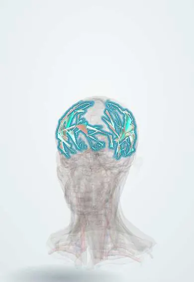
Representative model, for educational purposes only.