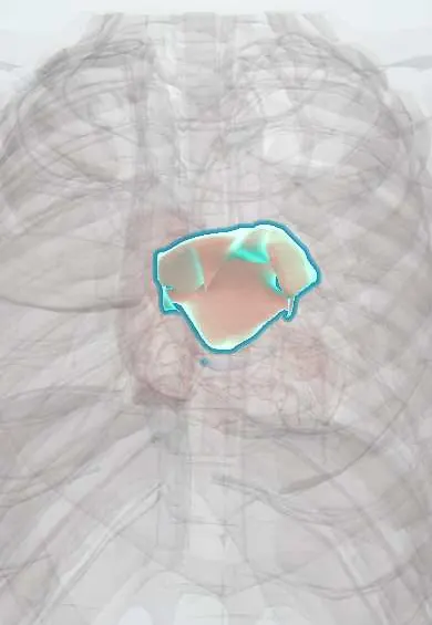
Atrial fibrillation !
Atrial fibrillation is the most common lasting irregular heartbeat: the upper chambers quiver instead of beating, raising the risk of clots and stroke.
Updated 2026-10-30
Structures involved
Tap one to see what it is and open it in 3D.
- Left atrium
- Right atrium
- Atrioventricular (AV) node
- Sinoatrial (SA) node
- Pulmonary veins
- Interatrial septum
- Left ventricle
- Bundle of His
In biochemistry
In histology
In embryology
In the clinical exam
- Jugular venous pressure and the venous pulse
- Abnormal pulses: paradoxus, alternans, parvus et tardus, bounding
- Acute limb ischemia and chronic arterial insufficiency
- Arterial pulse: rate, rhythm and amplitude
In physiology
- The cardiac conduction system
- The cardiac cycle
- ECG basics: waves, intervals and leads
- How arrhythmias arise
- Central venous pressure and the jugular pulse
Why it happens
- Arterial vs venous thrombi; mural thrombi and lines of Zahn
- Systemic (arterial) thromboembolism
- Thrombosis and Virchow's triad
- Alcohol: how it injures the liver, brain, heart and fetus
What it is
The most common sustained arrhythmia: disorganised electrical activity in the atria replaces normal sinus rhythm, so the atria quiver and the ventricles beat irregularly.
Who gets it
Becomes more common with age. In Latin America, rheumatic mitral disease and Chagas cardiomyopathy are important additional causes, often in younger patients. A 2016 review found that 46% of Latin American outpatients with atrial fibrillation lacked guideline-based anticoagulation, from 41.8% in Brazil to 54.8% in Colombia.
Causes
- Hypertension and hypertensive heart disease
- Heart failure and coronary artery disease
- Valve disease, especially rheumatic mitral stenosis
- Hyperthyroidism
- Alcohol ("holiday heart"), surgery, infection or pulmonary embolism
- Sometimes no cause is found
Risk factors
- Older age
- Hypertension
- Obesity and obstructive sleep apnoea
- Diabetes
- Heart failure
- Alcohol use
Pathophysiology
Rapid, chaotic impulses, often triggered from the pulmonary vein sleeves, bombard the atrioventricular node, which conducts them irregularly. The atria lose effective contraction, so blood stagnates, especially in the left atrial appendage, where clots form. Atrial stretch and fibrosis help the arrhythmia sustain itself.
Classification
- Paroxysmal: ends on its own or with treatment within 7 days
- Persistent: lasts more than 7 days
- Long-standing persistent: more than 1 year when rhythm control is chosen
- Permanent: patient and clinician accept the arrhythmia and stop trying to restore rhythm
- Valvular (moderate–severe mitral stenosis or mechanical valve) vs non-valvular
Symptoms and signs
- Palpitations
- Irregularly irregular pulse
- Breathlessness and reduced exercise tolerance
- Fatigue, dizziness
- Chest discomfort
- Can be silent and found by chance or after a stroke
Diagnosis
- Clinical
- Irregularly irregular pulse; pulse deficit between apex and radial pulse. Stroke risk is estimated with CHA2DS2-VASc and bleeding risk with tools such as HAS-BLED; guidelines vary on the scores used.
- Tests
- 12-lead ECG: no P waves, fibrillatory baseline and irregularly irregular RR intervals. Holter or event monitoring for paroxysmal episodes. Thyroid function, electrolytes, kidney function and blood count.
- Imaging
- Echocardiography for atrial size, valves (mitral stenosis) and ventricular function; transoesophageal echo looks for left atrial appendage clot before cardioversion.
Differential diagnosis
- Atrial flutter with variable block
- Multifocal atrial tachycardia
- Sinus rhythm with frequent ectopic beats
- Other supraventricular tachycardias
Goal of treatment
Prevent stroke and systemic embolism, control symptoms with rate or rhythm control, and treat the underlying causes.
Treatment
- Acute
- Unstable patients: urgent synchronised electrical cardioversion.
- Medical
- Rate control: beta-blockers or non-dihydropyridine calcium channel blockers, sometimes digoxin, to slow conduction through the atrioventricular node.
- Medical
- Anticoagulation according to stroke risk (CHA2DS2-VASc): direct oral anticoagulants, or vitamin K antagonists for moderate–severe mitral stenosis or mechanical valves.
- Procedure
- Rhythm control in selected patients: antiarrhythmic drugs, electrical cardioversion and catheter ablation (pulmonary vein isolation); left atrial appendage occlusion when anticoagulation is not possible.
- Lifestyle
- Control of blood pressure, weight, sleep apnoea and alcohol reduces recurrences.
Drugs for this condition
- Warfarin
- Rivaroxaban
- Dabigatran
- Metoprolol
- Digoxin
- Bisoprolol
- Diltiazem
- Esmolol
- Verapamil
- Sotalol
- Apixaban
- Edoxaban
Complications
- Ischaemic stroke and systemic embolism
- Tachycardia-induced cardiomyopathy and heart failure
- Bleeding related to anticoagulation
- Cognitive decline
Prognosis
Often recurrent and progressive, but with anticoagulation and good rate or rhythm control most patients live well. Stroke risk rises with the CHA2DS2-VASc score; untreated, it is a major cause of disabling stroke.
Prevention
- Control of hypertension, obesity and diabetes
- Moderate alcohol
- Treatment of sleep apnoea
- Prevention of rheumatic heart disease
Test yourself
Think of the answer, then tap the question to see it.
What are the ECG features of atrial fibrillation?
No P waves, a fibrillatory baseline and irregularly irregular RR intervals, usually with narrow QRS complexes.
What score estimates stroke risk in non-valvular atrial fibrillation?
CHA2DS2-VASc: heart failure, hypertension, age, diabetes, prior stroke/TIA, vascular disease and sex category.
Where do most clots form in atrial fibrillation?
In the left atrial appendage, where blood stagnates when the atria stop contracting effectively.
What is the first step in an unstable patient with fast atrial fibrillation?
Urgent synchronised electrical cardioversion.
Which anticoagulant class is used in AF with moderate–severe mitral stenosis or a mechanical valve?
Vitamin K antagonists; direct oral anticoagulants are not used in these settings.
Related conditions
- Heart failure
- Hypertensive heart disease
- Rheumatic heart disease
- Arterial hypertension
- Acute mesenteric ischaemia
- Ischaemic stroke
- Graves disease (hyperthyroidism)
- Transient ischaemic attack
Educational information, not medical advice. For any symptom, see a health professional.
Sources: Atrial Fibrillation (StatPearls (NCBI Bookshelf), 2026); Targeting stroke risk and improving outcomes in patients with atrial fibrillation in Latin America (Sao Paulo medical journal, 2016) · adapted, CC BY 4.0.
Spotted a mistake?We are always improving for the community. Tell us what is wrong and our team will review it.Report a mistake
Representative model, for educational purposes only.