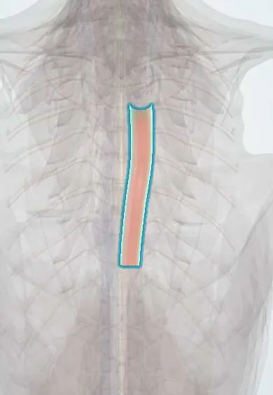
Thoracic aorta !
The thoracic aorta is the main artery running down the chest on the left front of the thoracic vertebrae; it feeds the body and the thoracic spine.
Opens in the Durgical app, no sign-up needed.
What it is
The main artery running down the chest on the left front of the thoracic vertebrae.
What it does
Carries blood to the body and gives off the posterior intercostal arteries that feed the thoracic spine.
Key facts
- Origin
- Continuation of the aortic arch at T4–T5; descends in posterior mediastinum left of the vertebral bodies to the aortic hiatus (T12)
- Branches
- Posterior intercostal arteries 3–11, subcostal; bronchial, oesophageal, pericardial, mediastinal, superior phrenic
- Supplies
- Thoracic wall, thoracic vertebrae and part of the cord, oesophagus, bronchi, posterior mediastinum
Relations
- Course
- From the end of the arch at T4–T5 it descends in the posterior mediastinum, drifting from the left side of the vertebral bodies toward the midline, and leaves the thorax behind the median arcuate ligament at T12
- In front
- Root of the left lung, the pericardium over the left atrium and, lower down, the oesophagus, which crosses in front of it toward the left
- Behind
- Thoracic vertebral bodies and the hemiazygos and accessory hemiazygos veins
- Medial
- On its right: the thoracic duct, the azygos vein and, in the upper part, the oesophagus
- Lateral
- Left mediastinal pleura and left lung
How to find it
It cannot be felt. In a left thoracotomy or thoracoscopy it runs down the left side of the vertebral bodies from the lower border of T4, drifting toward the midline, and leaves through the aortic hiatus in front of T12; a posterior intercostal artery crosses each vertebral body from it.
On imaging
On a frontal chest radiograph its left border makes a line beside the spine below the aortic knob; a widened mediastinum after trauma raises suspicion of aortic injury. CT angiography shows a dissection flap separating true and false lumens. Transesophageal echocardiography sees it well, because the oesophagus runs right beside it, first on its right and then in front of it.
Clinical notes
- Anterior approaches to the mid-thoracic spine are often made from the right to keep away from the aorta and heart; at the thoracolumbar junction the left is often preferred, because the aorta is easier to move than the vena cava and liver
- Left-sided thoracic pedicle screws that breach the front or side of the vertebral body can come to lie against the aorta, a known concern in scoliosis surgery
- A thoracic aortic aneurysm can erode the front of the adjacent vertebral bodies on imaging while the discs, which have no blood vessels, are spared
Anatomical variants
- With a right-sided aortic arch the descending aorta may run down the right side of the spine (rare)
- An aberrant right subclavian artery can arise from the start of the descending aorta and pass behind the oesophagus; most people have no symptoms, a few have trouble swallowing (dysphagia lusoria)
Development
Below about T4 it comes from the fused pair of dorsal aortae; above that level it comes from the left dorsal aorta alone. Coarctation usually lies at the isthmus, where the ductus arteriosus joins.
Related conditions
- Aortic dissection
- Takayasu arteritis
- Blunt traumatic aortic injury
- Thoracic aortic aneurysm and aortitis
- Coarctation of the aorta
Test yourself
Think of the answer, then tap the question to see it.
Which part of the aorta does a Stanford type B dissection involve, and what is the classic pain?
Only the descending aorta, beyond the left subclavian artery; tearing pain between the shoulder blades.
Why is paraplegia a key risk of thoracoabdominal aortic surgery?
The lower left posterior intercostal arteries usually give the artery of Adamkiewicz, which feeds the lower spinal cord.
Where does blunt deceleration injury tear the thoracic aorta?
At the isthmus, just beyond the left subclavian artery, where the ligamentum arteriosum tethers it.
Why does a thoracic aortic aneurysm erode vertebral bodies but spare the discs?
The discs have no blood vessels, so they resist the erosion that affects the adjacent vertebral bodies.
Educational information, not medical advice. For any symptom, see a health professional.
Its names
- Official Latin (TA2)
- aorta thoracica TA2 4186
- Official Spanish (DeCS)
- Aorta torácica DeCS D001013
- English (TA2)
- Thoracic aorta
- Also called
- descending thoracic aorta · Aorta, Thoracic · aorta thoracalis · aorta thoracica descendens
Related
Sources: Terminologia Anatomica (TA2); DeCS; Anatomy, Abdomen and Pelvis: Aorta; Anatomy of the Human Body (20th ed.): The Descending Aorta (Lea & Febiger (Gray's Anatomy, 1918), 1918) · adapted, Public domain; Anatomy of the Human Body (20th ed.): Development of the Vascular System (Lea & Febiger (Gray's Anatomy, 1918), 1918) · adapted, Public domain; Tratado de anatomía humana (9.ª ed.), tomo II, Libro IV, sección 2 Arterias, caps. I-III (sistema de la arteria pulmonar; aorta y ramas del cayado hasta la carótida interna).
Spotted a mistake?We are always improving for the community. Tell us what is wrong and our team will review it.Report a mistake
Representative model, for educational purposes only.