Atrial flutter !
Atrial flutter is a fast, regular rhythm caused by an electrical circuit spinning in the right atrium. Like atrial fibrillation, it raises the risk of stroke.
Updated 2026-11-01
Structures involved
Tap one to see what it is and open it in 3D.
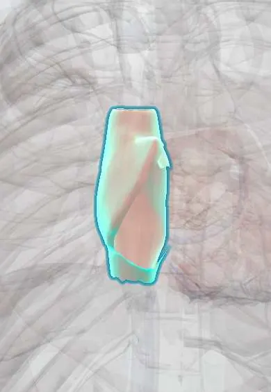
- Right atrium
- Tricuspid septal leaflet
- Coronary sinus
- Inferior vena cava
- Tricuspid anterior leaflet
- Tricuspid posterior leaflet
- Atrioventricular (AV) node
- Left atrium
- Interatrial septum
Why it happens
What it is
A macro-reentrant atrial tachycardia in which an electrical wave circles the right atrium at about 300 beats per minute. In typical flutter the circuit passes through the cavotricuspid isthmus, between the tricuspid valve and the inferior vena cava. The atrioventricular node usually conducts every second wave, giving a ventricular rate near 150 beats per minute.
Who gets it
Less common than atrial fibrillation, and often coexists with it. It increases with age and is more frequent in men and in people with heart or lung disease. In Colombia and Latin America, rheumatic valve disease, Chagas cardiomyopathy and the population with hypertension and chronic lung disease contribute to the burden.
Causes
- Structural heart disease: hypertension, heart failure, coronary disease, valve disease (especially mitral)
- Lung disease: chronic obstructive pulmonary disease, pulmonary embolism, pulmonary hypertension
- After cardiac surgery, including repair of congenital heart disease
- Hyperthyroidism, alcohol excess and acute illness
- Scar or prior ablation for atrial fibrillation (atypical flutter)
Risk factors
- Older age and male sex
- Hypertension and heart failure
- Chronic obstructive pulmonary disease and obstructive sleep apnoea
- Previous cardiac surgery
- Atrial fibrillation treated with antiarrhythmic drugs
Pathophysiology
A slow-conduction zone at the cavotricuspid isthmus allows a single wave to travel around the tricuspid annulus, usually counterclockwise. Because the circuit is large and stable, atrial activity is regular. The atrioventricular node filters impulses, typically 2:1, so the pulse is regular and fast. Blood stagnates in the atria, especially the left atrial appendage, favouring thrombus formation and embolic stroke, much as in atrial fibrillation.
Classification
- Typical (cavotricuspid isthmus-dependent): counterclockwise (most common) or clockwise
- Atypical (non-isthmus-dependent): circuits around surgical scars or in the left atrium
- By atrioventricular conduction ratio: 2:1 (most common), 3:1, 4:1 or variable
Symptoms and signs
- Palpitations with a fast, regular pulse
- Dyspnoea, fatigue and reduced exercise tolerance
- Dizziness, chest discomfort or syncope
- Worsening heart failure in patients with heart disease
- Sometimes no symptoms
Diagnosis
- Clinical
- Suspect it in a patient with a regular tachycardia around 150 beats per minute that does not vary with activity. Assess haemodynamic stability first.
- Tests
- 12-lead ECG: saw-tooth flutter waves without an isoelectric baseline, best seen in the inferior leads (II, III, aVF) and V1, with regular QRS complexes. Vagal manoeuvres or adenosine slow the ventricular response transiently and unmask the flutter waves, without terminating the circuit. Thyroid function, electrolytes and kidney function.
- Imaging
- Transthoracic echocardiography to assess atrial size, valves and ventricular function; transoesophageal echocardiography to rule out atrial thrombus before cardioversion when anticoagulation has not been adequate.
Differential diagnosis
- Sinus tachycardia
- Atrial fibrillation (irregular rhythm without organised atrial waves)
- Atrioventricular nodal re-entrant tachycardia
- Focal atrial tachycardia
- Ventricular tachycardia when there is bundle branch block
Goal of treatment
Control the ventricular rate, restore sinus rhythm, prevent stroke with anticoagulation according to stroke risk, and cure typical flutter with ablation of the cavotricuspid isthmus.
Treatment
- Acute
- Unstable patient: synchronised electrical cardioversion; flutter usually responds to low energy.
- Medical
- Rate control in stable patients with beta-blockers or non-dihydropyridine calcium channel blockers; rate control is often harder than in atrial fibrillation.
- Medical
- Anticoagulation following the same stroke-risk criteria as for atrial fibrillation, including before and after cardioversion.
- Procedure
- Catheter ablation of the cavotricuspid isthmus: first-line long-term treatment for typical flutter, with a high cure rate.
- Medical
- Antiarrhythmic drugs or elective cardioversion when ablation is not chosen; class IC drugs are given with an atrioventricular nodal blocker to avoid 1:1 conduction.
- Follow-up
- Nursing priorities: continuous ECG monitoring, blood pressure, symptoms of low output, neurological checks and safety of anticoagulation (bleeding signs).
Complications
- Embolic stroke and systemic embolism
- Tachycardia-induced cardiomyopathy and heart failure
- Progression to atrial fibrillation
- 1:1 conduction with very fast rates and collapse
Prognosis
Typical flutter is usually cured by isthmus ablation, although many patients later develop atrial fibrillation. Outcome depends largely on the underlying heart or lung disease and on preventing stroke.
Prevention
- Control of hypertension, heart failure and lung disease
- Moderation of alcohol and treatment of sleep apnoea and hyperthyroidism
- Anticoagulation according to stroke risk in patients with flutter
Test yourself
Think of the answer, then tap the question to see it.
What is the typical ECG appearance of atrial flutter?
Saw-tooth flutter waves at about 300 per minute, best seen in II, III, aVF and V1, usually with 2:1 conduction.
A regular narrow-complex tachycardia fixed at 150 per minute should make you think of what?
Atrial flutter with 2:1 atrioventricular block.
Where is the critical isthmus of typical flutter located?
In the right atrium, between the tricuspid valve annulus and the inferior vena cava (cavotricuspid isthmus).
What is the long-term treatment of choice for typical atrial flutter?
Catheter ablation of the cavotricuspid isthmus.
Does atrial flutter require stroke prevention?
Yes. Anticoagulation follows the same stroke-risk criteria as atrial fibrillation.
Related conditions
- Atrial fibrillation
- Ischaemic stroke
- Heart failure
- Supraventricular tachycardia (AVNRT, AVRT)
- Graves disease (hyperthyroidism)
- Chronic obstructive pulmonary disease (COPD)
- Obstructive sleep apnoea
- Pulmonary embolism
- Mitral stenosis
Educational information, not medical advice. For any symptom, see a health professional.
Sources: Atrial fibrillation and atrial flutter: MedlinePlus Medical Encyclopedia (MedlinePlus, US National Library of Medicine, 2026).
Spotted a mistake?We are always improving for the community. Tell us what is wrong and our team will review it.Report a mistake
Representative model, for educational purposes only.