Glioblastoma !
Glioblastoma is the most common and aggressive malignant brain tumour in adults; it grows fast and spreads into nearby brain tissue.
Updated 2026-10-29
Structures involved
Tap one to see what it is and open it in 3D.
- White matter
- Frontal lobe
- Temporal lobe
- Corpus callosum
- Parietal lobe
- Lateral ventricles
- Middle cerebral artery
- Motor cortex
In biochemistry
- Isocitrate dehydrogenase
- Oncogenes and tumour suppressor genes
- Warburg effect (aerobic glycolysis in cancer)
In histology
In the clinical exam
Why it happens
- Tumour angiogenesis
- Oncogenes: RAS, MYC, HER2, BCR-ABL and the table to know
- Differentiation and anaplasia: how a tumour cell looks under the microscope
- Translocations, amplifications and epigenetic changes in cancer
- Benign vs malignant tumours: the comparison table
What it is
The most common and most aggressive primary malignant brain tumour in adults, arising from glial cells. It grows fast and infiltrates surrounding brain, so it is hard to remove completely.
Who gets it
Usually affects adults over 50, slightly more often men. It is the most frequent malignant primary brain tumour; most cases arise de novo without a known cause.
Causes
- Mostly sporadic, from accumulated genetic changes in glial cells
- Prior therapeutic radiation to the head (established risk factor)
- Rare inherited syndromes (e.g. Li-Fraumeni, neurofibromatosis type 1)
Risk factors
- Older age
- Male sex
- Previous cranial radiotherapy
- Inherited cancer syndromes
Pathophysiology
Under the current WHO classification, glioblastoma is an IDH-wildtype diffuse astrocytic tumour of grade 4. It shows necrosis, microvascular proliferation and tumour cells that infiltrate along white matter tracts, sometimes crossing the corpus callosum (butterfly glioma). Mass effect, oedema and seizures arise from local invasion.
Symptoms and signs
- Progressive headache, often worse in the morning
- Seizures
- Progressive focal deficits: weakness, speech or visual disturbance
- Personality change, confusion, memory problems
- Nausea and vomiting from raised intracranial pressure
Diagnosis
- Clinical
- Subacute progressive neurological symptoms in an adult, with signs of raised intracranial pressure or focal deficit on examination.
- Tests
- Tissue diagnosis by biopsy or resection with histology and molecular markers (IDH status, MGMT promoter methylation, others), which guide classification and prognosis.
- Imaging
- MRI with gadolinium: irregular ring-enhancing mass with central necrosis and surrounding oedema, often crossing the corpus callosum. Perfusion MRI and spectroscopy help distinguish it from other lesions.
Differential diagnosis
- Brain metastasis
- Brain abscess
- Primary central nervous system lymphoma
- Neurocysticercosis or tuberculoma
- Tumefactive demyelination
- Lower-grade glioma
Goal of treatment
Obtain a diagnosis, remove as much tumour as safely possible, control growth and symptoms and preserve quality of life.
Treatment
- Surgical
- Maximal safe resection, or biopsy when the tumour is in an eloquent or deep area.
- Medical
- Radiotherapy with concurrent and then adjuvant alkylating chemotherapy is the standard after surgery in fit patients.
- Medical
- Corticosteroids for oedema and antiseizure drugs when seizures occur.
- Procedure
- Tumour-treating fields are offered in some centres alongside chemotherapy.
- Follow-up
- Regular MRI follow-up, palliative care involvement and support for patient and family.
Complications
- Seizures
- Raised intracranial pressure and herniation
- Progressive neurological disability
- Venous thromboembolism
- Adverse effects of corticosteroids, chemotherapy and radiotherapy
Prognosis
Poor: recurrence is almost universal and median survival with standard treatment is measured in months to under two years. Younger age, good functional status, more complete resection and MGMT promoter methylation are linked with longer survival.
Test yourself
Think of the answer, then tap the question to see it.
What are the typical MRI findings of glioblastoma?
An irregular ring-enhancing mass with central necrosis and surrounding oedema, sometimes crossing the corpus callosum (butterfly glioma).
Which molecular marker predicts better response to alkylating chemotherapy in glioblastoma?
Methylation of the MGMT promoter.
In the current WHO classification, what IDH status defines glioblastoma?
IDH-wildtype; IDH-mutant grade 4 tumours are classified as astrocytomas.
What is the standard treatment after maximal safe resection of glioblastoma?
Radiotherapy with concurrent and adjuvant alkylating chemotherapy.
Related conditions
Educational information, not medical advice. For any symptom, see a health professional.
Sources: Glioblastoma Multiforme (StatPearls (NCBI Bookshelf), 2024).
Spotted a mistake?We are always improving for the community. Tell us what is wrong and our team will review it.Report a mistake
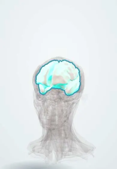
Representative model, for educational purposes only.