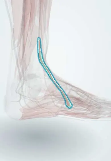
Fibularis tertius !
The fibularis tertius is a small muscle at the front of the leg reaching the fifth metatarsal; it helps lift the foot and turn it outward.
Official name: Fibularis tertius muscle
Opens in the Durgical app, no sign-up needed.
What it is
A small muscle on the front of the leg, often joined to the extensor digitorum longus. Its tendon goes to the fifth metatarsal. Not everyone has it.
What it does
Helps lift the foot and turn it outward.
Key facts
- Origin
- Lower third of anterior fibula; interosseous membrane (part of extensor digitorum longus; sometimes absent)
- Insertion
- Dorsal base of fifth metatarsal
- Action
- Dorsiflexes ankle; everts foot
- Nerve
- Deep fibular nerve (L5–S1)
- Blood supply
- Anterior tibial artery
Relations
- Medial
- Extensor digitorum longus tendons, with which it shares a synovial sheath under the extensor retinacula
- Lateral
- Lateral malleolus; in the leg, fibularis brevis beyond the anterior intermuscular septum
- Below
- Dorsal base of the fifth metatarsal, next to the fibularis brevis insertion on the tuberosity
- Superficial
- Inferior extensor retinaculum, branches of the superficial fibular nerve and the skin of the lateral dorsum
How to find it
With the foot lifted and turned out, a thin tendon may be felt lateral to the extensor digitorum longus tendon for the little toe, heading for the base of the fifth metatarsal; when the muscle is absent no such tendon is found.
On imaging
Usually not imaged on its own. On ultrasound or axial MRI it is the most lateral tendon in front of the ankle, lateral to the extensor digitorum longus, ending on the dorsal base of the fifth metatarsal. Its absence in some people is a normal variant, not a tear. Foot radiographs are checked for a fracture of the fifth metatarsal base nearby.
Clinical notes
- Because it shares the deep fibular nerve with the other dorsiflexors, it cannot make up for foot drop caused by that nerve
- Its tendon is the lateral landmark for anterior ankle arthroscopy: the anterolateral portal goes just lateral to it, where branches of the superficial fibular nerve are at risk
Anatomical variants
- May be absent or small; how often varies between populations
- It may be fused so closely with extensor digitorum longus that it does not look like a separate muscle
Memory aid
The last of "Tom Has Very Nice Dancing Pigs" in front of the ankle: Pigs = Peroneus tertius.
Test yourself
Think of the answer, then tap the question to see it.
Which compartment does the fibularis tertius belong to, and what is its nerve supply?
The anterior compartment; deep fibular nerve, not the superficial one.
Where does the fibularis tertius insert, and what are its actions?
On the dorsal base of the fifth metatarsal; it dorsiflexes the ankle and everts the foot.
Why can't the fibularis tertius compensate for foot drop from a deep fibular nerve lesion?
It shares the deep fibular nerve with the other dorsiflexors, so it is weak too.
Educational information, not medical advice. For any symptom, see a health professional.
Its names
- Official Latin (TA2)
- musculus fibularis tertius TA2 2649
- Official Spanish (DeCS)
- Músculo fibular tercero
- English (TA2)
- Fibularis tertius muscle
- Also called
- musculus peroneus tertius · peroneus tertius muscle
Related
- Extensor digitorum longus
- Fifth metatarsal
- Common fibular nerve
- Deep fibular nerve
- Anterior tibial artery
- Fibular artery
- Fibula
- Tibia
Sources: Terminologia Anatomica (TA2); Anatomy, Bony Pelvis and Lower Limb: Leg Anterior Compartment.
Spotted a mistake?We are always improving for the community. Tell us what is wrong and our team will review it.Report a mistake
Representative model, for educational purposes only.