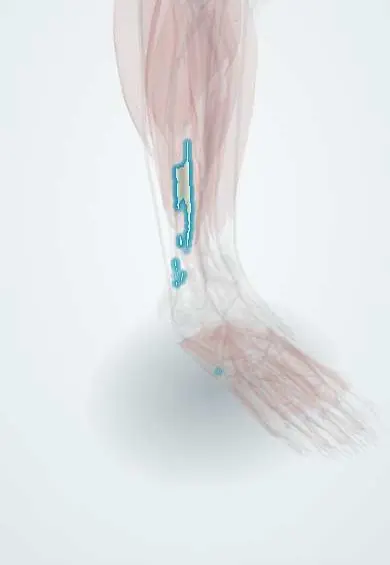
Fibularis brevis !
The fibularis brevis is a short muscle on the outer side of the leg, under the fibularis longus; it steadies the ankle.
Official name: Fibularis brevis muscle
Opens in the Durgical app, no sign-up needed.
What it is
A short muscle on the outer side of the leg.
What it does
Turns the foot outward.
Key facts
- Origin
- Lower two-thirds of lateral surface of fibula
- Insertion
- Tuberosity on base of fifth metatarsal
- Action
- Everts foot; weak plantar flexion
- Nerve
- Superficial fibular nerve (L5–S2)
- Blood supply
- Fibular artery; anterior tibial artery
Relations
- Course
- From the lower fibula its tendon grooves the back of the lateral malleolus, passes above the fibular trochlea of the calcaneus (longus passes below) and reaches the fifth metatarsal tuberosity
- Superficial
- Fibularis longus in the leg and behind the malleolus; the superior and inferior fibular retinacula
- Deep
- Fibula; at the ankle, the calcaneofibular ligament lies deep to both fibular tendons
- In front
- Extensor digitorum longus and fibularis tertius, beyond the anterior intermuscular septum
- Behind
- Flexor hallucis longus, beyond the posterior intermuscular septum
How to find it
With the foot everted, its tendon is felt from behind the lateral malleolus to the prominent base of the fifth metatarsal, where it stands out just behind the tuberosity.
Examination
- Resisted eversion with the ankle plantarflexed: pain along the tendon behind the malleolus suggests tendinopathy or a split tear; weakness suggests a superficial fibular nerve or root problem
- Fibular tendon subluxation test: snapping of the tendons over the malleolus during resisted eversion and dorsiflexion
On imaging
Foot radiographs show where a fifth metatarsal base fracture lies: tuberosity avulsion versus the junction of the metaphysis and diaphysis. On MRI or ultrasound a split tear looks like a flattened or divided tendon behind the malleolus.
Clinical notes
- Split tears often follow repeated ankle sprains and accompany lateral ankle instability
- Its tendon has been used to reconstruct the lateral ankle ligaments, although anatomical repairs are now favoured
- A low-lying muscle belly or a fibularis quartus can crowd the retromalleolar groove and predispose to tendon problems
Anatomical variants
- Fibularis quartus, an extra muscle from the lower fibula to the calcaneus, is reported in about 5–20% of legs
- The retromalleolar groove is concave in most people; a flat or convex groove favours tendon dislocation
- Its muscle belly may extend lower than usual into the groove
Development
A dorsal muscle mass derivative of the lateral compartment, supplied by the superficial fibular nerve.
Related conditions
Test yourself
Think of the answer, then tap the question to see it.
Where does the fibularis brevis insert, and what fracture can an inversion injury cause there?
On the tuberosity of the fifth metatarsal base; an inversion injury can avulse this tuberosity, which differs from the more distal Jones fracture.
Which fibular tendon usually develops longitudinal split tears, and why?
The fibularis brevis, because it lies directly against the back of the lateral malleolus, squeezed between bone and the fibularis longus.
Which nerve supplies the fibularis brevis?
The superficial fibular nerve (L5–S2), as for the fibularis longus in the lateral compartment.
Educational information, not medical advice. For any symptom, see a health professional.
Its names
- Official Latin (TA2)
- musculus fibularis brevis TA2 2653
- Official Spanish (DeCS)
- Músculo fibular corto
- English (TA2)
- Fibularis brevis muscle
- Also called
- musculus peroneus brevis · peroneus brevis muscle
Related
- Common fibular nerve
- Superficial fibular nerve
- Fibular artery
- Fibularis brevis tendon
- Fibula
- Fifth metatarsal
Sources: Terminologia Anatomica (TA2); Anatomy, Bony Pelvis and Lower Limb, Foot Peroneus Brevis Muscle.
Spotted a mistake?We are always improving for the community. Tell us what is wrong and our team will review it.Report a mistake
Representative model, for educational purposes only.