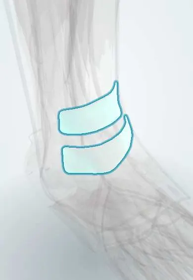
Extensor retinacula !
The extensor retinacula are bands across the front of the ankle that hold the front tendons close to the bone.
Official name: Superior extensor retinaculum of ankle
Opens in the Durgical app, no sign-up needed.
What it is
Two bands across the front of the ankle: one above the joint and a Y-shaped one below it.
What it does
Hold the front tendons close to the bones so they pull efficiently when the ankle bends.
Key facts
- Attaches
- Superior: anterior borders of tibia and fibula above ankle; inferior (Y-shaped): calcaneus to medial malleolus and plantar fascia
- Function
- Hold tibialis anterior, EHL, EDL and fibularis tertius tendons to ankle; prevent bowstringing
Relations
- Contents
- Medial to lateral: tibialis anterior tendon, extensor hallucis longus, anterior tibial artery (becoming the dorsalis pedis artery) with its veins and the deep fibular nerve, toe extensor tendons and fibularis tertius
- Superficial
- Branches of the superficial fibular nerve crossing over them; medially, the great saphenous vein and saphenous nerve in front of the medial malleolus
- Deep
- Ankle joint capsule, talar neck and the tendon sheaths
- Lateral
- The stem of the inferior retinaculum anchors in the sinus tarsi and continues into the inferior fibular retinaculum
How to find it
The superior band lies a few centimetres above the ankle joint line between the tibia and fibula; the inferior band crosses the front of the ankle and top of the talus. When the foot and toes are lifted, the tendons bulge between and beyond the bands.
Examination
- Tinel sign over the deep fibular nerve under the inferior retinaculum: tingling into the first web space suggests anterior tarsal tunnel syndrome
On imaging
Ultrasound and MRI show the superior and inferior extensor retinacula as thin dark bands across the extensor tendons in front of the ankle. Look for thickening or a tear after a sprain and sheath fluid beneath. In anterior tarsal tunnel syndrome, imaging looks for what presses on the deep fibular nerve, such as a dorsal talonavicular osteophyte or a ganglion; radiographs show the spurs.
Common problems
- Tight shoes or laces over the lower band can press on the deep fibular nerve (anterior tarsal tunnel).
Clinical notes
- Anterior tarsal tunnel syndrome: numbness in the first web space and, in long-standing cases, wasting of the extensor digitorum brevis; causes include tight laces, osteophytes and ganglia
- If the retinaculum is torn or released, the tibialis anterior tendon can bowstring across the front of the ankle
- Anterior ankle arthroscopy portals sit medial to the tibialis anterior and lateral to the fibularis tertius; the lateral portal is close to the superficial fibular nerve
Anatomical variants
- Some texts describe an extra upper-lateral band of the inferior retinaculum, so that it looks X-shaped rather than Y-shaped
Memory aid
Under the extensor retinacula, medial to lateral: "The Hospitals Are Not Dirty Places" (Tibialis anterior, Hallucis longus, Artery, Nerve, Digitorum longus, Peroneus tertius).
Test yourself
Think of the answer, then tap the question to see it.
What is the shape of the inferior extensor retinaculum, and where are its limbs attached?
Y-shaped: the stem anchors in the sinus tarsi and calcaneus, an upper limb reaches the medial malleolus and a lower limb blends with the plantar fascia.
What is anterior tarsal tunnel syndrome?
Compression of the deep fibular nerve under the inferior extensor retinaculum, giving numbness in the first web space.
What happens to the tibialis anterior tendon if the extensor retinaculum is torn or released?
It can bowstring across the front of the ankle.
Which nerve is close to the lateral portal of anterior ankle arthroscopy?
The superficial fibular nerve; the lateral portal sits lateral to the fibularis tertius.
Educational information, not medical advice. For any symptom, see a health professional.
Its names
- Official Latin (TA2)
- retinaculum extensorium superius tali; retinaculum extensorium inferius tali TA2 2712
- Official Spanish (DeCS)
- Retináculos extensores superior e inferior del tobillo
- English (TA2)
- Superior extensor retinaculum of ankle
- Also called
- transverse crural ligament · cruciate crural ligament · ligamentum transversum cruris
Related
- Tibialis anterior tendon
- Toe extensor tendon
- Extensor hallucis longus
- Deep fibular nerve
- Crural fascia
- Fibula
- Tibia
Sources: Terminologia Anatomica (TA2); Anatomy, Bony Pelvis and Lower Limb: Leg Anterior Compartment.
Spotted a mistake?We are always improving for the community. Tell us what is wrong and our team will review it.Report a mistake
Representative model, for educational purposes only.