Joints and ligaments · Upper limb
Pisiform ligaments !
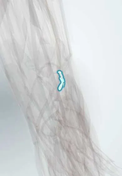
The pisiform ligaments tie the pisiform to the hamate and fifth metacarpal; they carry a wrist flexor's pull and floor Guyon's canal.
Official name: Pisohamate and pisometacarpal ligaments
Opens in the Durgical app, no sign-up needed.
What it is
Short bands that anchor the pisiform to the hamate and the 5th metacarpal.
What it does
Pass the pull of flexor carpi ulnaris to the hand; they form the floor of Guyon's canal.
Key facts
- Attaches
- Pisiform to the hook of hamate (pisohamate) and to the base of the 5th metacarpal (pisometacarpal)
- Function
- Carry the flexor carpi ulnaris pull on to the hand; pisohamate ligament is the floor of Guyon's canal
- Tight in
- Wrist extension; flexor carpi ulnaris contraction
Relations
- Superficial
- Guyon canal: ulnar nerve and ulnar artery with their veins, under the palmar carpal ligament and palmaris brevis
- Lateral
- Hook of the hamate, around which the deep branch of the ulnar nerve and the deep branch of the ulnar artery turn into the palm
- Deep
- Pisotriquetral joint and the triquetrum
- Below
- Origins of the hypothenar muscles: abductor digiti minimi from the pisiform, flexor digiti minimi and opponens digiti minimi from the hook of the hamate
How to find it
The pisiform is the pea-sized bump at the base of the hypothenar eminence, at the distal wrist crease, where flexor carpi ulnaris ends; it slides side to side with the wrist relaxed in slight flexion. The hook of the hamate is felt about 2 cm distal and radial to it, and the pisohamate ligament spans the gap.
Examination
- Tinel sign over Guyon canal: tapping between the pisiform and the hook of the hamate sends tingling into the little finger and the ulnar half of the ring finger, suggesting ulnar nerve irritation there
- Pisotriquetral grind test: the pisiform is pressed against the triquetrum and moved; pain or crepitus points to pisotriquetral arthritis or instability
- Froment sign: gripping paper between thumb and index, the thumb IP joint flexes as FPL substitutes for a weak adductor pollicis; seen with ulnar motor loss, including deep branch compression here
On imaging
The pisiform and the pisotriquetral joint are best seen on a lateral or semi-supinated oblique wrist view, since the PA view projects the pisiform over the triquetrum. The ligaments are seen on MRI or ultrasound, which also show Guyon canal with the ulnar artery and ulnar nerve beside the pisiform. A carpal tunnel view or CT shows the hook of the hamate.
Clinical notes
- Ulnar tunnel zones: zone 1 before the nerve divides (motor and sensory signs), zone 2 around the deep motor branch (weakness only), zone 3 around the superficial branch (sensory only)
- Causes include ganglia, hook of hamate fractures, ulnar artery thrombosis (hypothenar hammer syndrome) and repeated pressure from handlebars or tools
- Pisiform excision for painful pisotriquetral arthritis is done by shelling the bone out of the FCU tendon, keeping the tendon in continuity with these ligaments and guarding the ulnar nerve and artery in Guyon canal
Test yourself
Think of the answer, then tap the question to see it.
Where do the pisiform ligaments attach?
Pisohamate: pisiform to the hook of the hamate; pisometacarpal: pisiform to the base of the 5th metacarpal.
What do the pisiform ligaments transmit?
The pull of flexor carpi ulnaris, in which the pisiform is a sesamoid, on to the hamate and 5th metacarpal.
Which canal has the pisohamate ligament in its floor?
Guyon canal, with the flexor retinaculum; it carries the ulnar nerve and artery between the pisiform and the hook of the hamate.
Educational information, not medical advice. For any symptom, see a health professional.
Its names
- Official Latin (TA2)
- ligamentum pisohamatum; ligamentum ulnopisiforme TA2 1825
- Official Spanish (DeCS)
- Ligamentos pisiganchoso, pisometacarpiano y pisopiramidal
- English (TA2)
- Pisohamate and pisometacarpal ligaments
- Also called
- ulnopisiform ligament
Related
Sources: Terminologia Anatomica (TA2); Anatomy, Shoulder and Upper Limb, Hand Carpal Bones; Anatomy, Shoulder and Upper Limb, Wrist Joint.
Spotted a mistake?We are always improving for the community. Tell us what is wrong and our team will review it.Report a mistake
Representative model, for educational purposes only.