Joints and ligaments · Upper limb
Interosseous membrane !
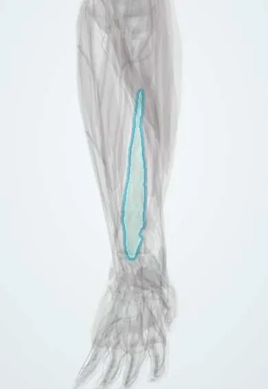
The interosseous membrane is a strong sheet joining the radius and ulna along the forearm; it ties them together and passes load between them.
Official name: Interosseous membrane of the forearm
Opens in the Durgical app, no sign-up needed.
What it is
A tough sheet between the radius and the ulna along the forearm.
What it does
Ties the two bones together and passes load from the radius to the ulna.
Key facts
- Attaches
- Interosseous borders of the radius and ulna (fibres run down and medially from radius to ulna)
- Function
- Transfers load from the radius to the ulna; separates the compartments; tightest in mid-prone position
Relations
- In front
- Deep flexors: flexor digitorum profundus (FDP), flexor pollicis longus (FPL) and, distally, pronator quadratus; the anterior interosseous nerve and artery lie directly on it
- Behind
- Deep extensors: abductor pollicis longus (APL), extensor pollicis brevis (EPB), extensor pollicis longus (EPL) and extensor indicis, with the posterior interosseous nerve and artery
- Above
- A gap between its upper edge and the oblique cord, below the radial tuberosity, through which the posterior interosseous vessels pass
- Below
- Ends just above the distal radioulnar joint, deep to pronator quadratus
How to find it
It cannot be felt. In dissection it appears once the deep flexors or deep extensors are lifted off the bones; on ultrasound it is a thin bright line bridging the radius and ulna on a transverse scan of the mid-forearm.
Examination
- Radius pull test (during surgery, with the radial head fractured or removed): the radius is pulled along its axis; proximal migration beyond a few millimetres suggests a torn membrane, and more still suggests TFCC injury as well
On imaging
On X-ray the clue to a membrane tear is indirect: a radial head fracture with the radius shortened at the wrist (positive ulnar variance). Ultrasound and MRI can show the central band torn or lax.
Clinical notes
- In an Essex-Lopresti injury, treating only the radial head fracture (or excising the radial head) lets the radius migrate proximally, with ulnar-sided wrist pain
- After forearm shaft fractures, malunion, scarring of the membrane or a bony bridge across it (radioulnar synostosis) limits pronation and supination
- In chronic longitudinal instability the central band can be rebuilt with a tendon graft or a suture construct, usually together with restoring the radial head
Test yourself
Think of the answer, then tap the question to see it.
Which way do the fibres of the forearm interosseous membrane run, and what does that achieve?
Down and medially from radius to ulna, transferring load from the radius to the ulna.
What is the Essex-Lopresti injury?
A radial head fracture with a torn interosseous membrane and a disrupted distal radioulnar joint, so the radius slides proximally.
What does the forearm interosseous membrane separate, and what runs on its two faces?
The anterior (flexor) and posterior (extensor) compartments; the anterior and posterior interosseous nerves and arteries run on its faces.
Why is excising the radial head alone harmful in an Essex-Lopresti injury?
With the interosseous membrane torn, the radius migrates proximally, causing ulnar-sided wrist pain.
Educational information, not medical advice. For any symptom, see a health professional.
Its names
- Official Latin (TA2)
- membrana interossea antebrachii TA2 1761
- Official Spanish (DeCS)
- Membrana interósea del antebrazo
- English (TA2)
- Interosseous membrane of the forearm
Related
Sources: Terminologia Anatomica (TA2); Anatomy, Shoulder and Upper Limb, Forearm Bones; Anatomy, Shoulder and Upper Limb, Proximal Radio-Ulnar Joint; Anatomía humana (4.ª ed.), tomo 1, cap. 57: Antebrazo. Aparato de la pronosupinación.
Spotted a mistake?We are always improving for the community. Tell us what is wrong and our team will review it.Report a mistake
Representative model, for educational purposes only.