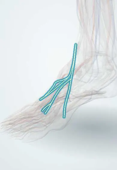
Top-of-foot skin nerves !
The top-of-foot skin nerves, mainly from the superficial fibular nerve, carry feeling from the top of the foot.
Official name: Medial dorsal cutaneous nerve of foot
Opens in the Durgical app, no sign-up needed.
What it is
Skin branches of the superficial fibular nerve on the top of the foot.
What it does
Carry feeling from most of the top of the foot and toes.
Key facts
- Roots
- L4–S1 (medial and intermediate dorsal cutaneous branches of superficial fibular nerve)
- Moves
- None (purely sensory, cutaneous)
- Feeling from
- Dorsum of foot and toes except first web space (deep fibular) and lateral border (sural)
Relations
- Course
- The medial dorsal cutaneous nerve crosses the front of the ankle to the medial big toe and the second web space; the intermediate one runs over the dorsum to the third and fourth web spaces; the lateral dorsal cutaneous nerve follows the lateral border of the foot to the little toe
- Superficial
- Skin only
- Deep
- The extensor retinaculum, the extensor hallucis longus and extensor digitorum longus tendons and the dorsal veins; deeper still, the deep fibular nerve and the dorsalis pedis artery
How to find it
With the ankle plantar-flexed and the foot inverted, the branches can often be seen or rolled under the fingertip over the front of the ankle and the dorsum in slim people, especially the intermediate branch heading to the fourth toe.
Examination
- Light touch on the dorsum: a numb patch that spares the first web space and the lateral border fits these branches; tapping over a scar or a tight shoe strap reproduces tingling
On imaging
Usually not seen on radiographs or routine MRI. High-resolution ultrasound can follow the superficial fibular branches in the fat over the extensor tendons and find a neuroma or scar tethering after a cut or surgery. The deep fibular nerve is found next to the dorsalis pedis artery, a useful landmark on Doppler ultrasound. Ultrasound can also guide a nerve block.
Common problems
- Can be irritated by tight shoes or straps, or injured during ankle and foot surgery, causing numbness or tingling.
Clinical notes
- At risk in anterolateral ankle arthroscopy portals and dorsal foot incisions such as midfoot fixation; the dorsomedial nerve of the big toe, a terminal branch, is the one hurt in bunion surgery
- Compressed by dorsal osteophytes over the midfoot joints or by a ganglion
- Blocked by a ring of subcutaneous local anaesthetic across the front of the ankle in an ankle block
Anatomical variants
- The split of the dorsum between the sural nerve and the superficial fibular nerve varies: the lateral dorsal cutaneous nerve may supply only the little toe or reach the fourth toe, with the intermediate dorsal cutaneous nerve taking the rest
Test yourself
Think of the answer, then tap the question to see it.
Which nerves supply the skin of the dorsum of the foot?
Medial and intermediate dorsal cutaneous nerves (superficial fibular) and the lateral dorsal cutaneous nerve (sural); the first web space is the deep fibular nerve's.
Why are the dorsal foot skin nerves so often injured in surgery?
They lie just under the skin over the extensor tendons, so incisions, portals and pins on the top of the foot reach them.
Which terminal dorsal nerve is the one injured in bunion surgery?
The dorsomedial nerve of the big toe, a terminal branch of the medial dorsal cutaneous nerve.
Educational information, not medical advice. For any symptom, see a health professional.
Its names
- Official Latin (TA2)
- nervus cutaneus medialis dorsalis pedis; nervus cutaneus intermedius dorsalis pedis; nervus cutaneus lateralis dorsalis pedis TA2 6576
- Official Spanish (DeCS)
- Nervios cutáneos dorsales medial, intermedio y lateral del pie
- English (TA2)
- Medial dorsal cutaneous nerve of foot
- Also called
- medial cutaneous nerve of dorsum of foot · intermediate cutaneous nerve of dorsum of foot · lateral cutaneous nerve of dorsum of foot · nervus cutaneus dorsi pedis tibialis
Related
Sources: Terminologia Anatomica (TA2); Anatomy, Bony Pelvis and Lower Limb, Foot Nerves.
Spotted a mistake?We are always improving for the community. Tell us what is wrong and our team will review it.Report a mistake
Representative model, for educational purposes only.