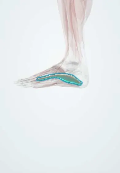
Abductor hallucis !
The abductor hallucis runs along the inner edge of the sole; it moves the big toe away from the others and supports the arch.
Opens in the Durgical app, no sign-up needed.
What it is
A muscle along the inner edge of the sole, from the heel bone to the big toe.
What it does
Moves the big toe away from the other toes and helps support the inner arch.
Key facts
- Origin
- Medial process of calcaneal tuberosity; flexor retinaculum; plantar fascia
- Insertion
- Medial base of proximal phalanx of big toe (with medial flexor hallucis brevis tendon)
- Action
- Abducts and flexes big toe; supports medial longitudinal arch
- Nerve
- Medial plantar nerve (S1–S2) (some texts: S2–S3)
- Blood supply
- Medial plantar artery
Relations
- Above
- Flexor retinaculum and the outlet of the tarsal tunnel, at its origin
- Deep
- Medial and lateral plantar nerves and vessels, the flexor hallucis longus and flexor digitorum longus tendons, quadratus plantae and, further forward, flexor hallucis brevis
- Lateral
- Flexor digitorum brevis, with the medial plantar nerve and vessels emerging in the groove between them
- Superficial
- Medial part of the plantar fascia and the skin of the medial arch
How to find it
It forms the soft bulge along the inner border of the foot under the medial arch, from the heel to the big toe; it tightens when the big toe is moved away from the others, a movement many people find hard to make.
Examination
- Tinel sign over the tarsal tunnel and the upper border of the muscle: tingling into the sole suggests compression of the tibial nerve or its plantar branches
On imaging
Ultrasound and MRI show it along the medial border of the foot, with the plantar nerves and vessels passing deep to it as they leave the tarsal tunnel. In tarsal tunnel syndrome, imaging looks for a space-occupying cause there, such as a ganglion, varicose veins or an accessory muscle. Denervation oedema or fatty atrophy of this muscle on MRI points to medial plantar nerve involvement.
Common problems
- Like the other small muscles of the foot, it can weaken with age, nerve problems or tight shoes, which can let the arch drop or the toes change shape.
Clinical notes
- "Jogger's foot": the medial plantar nerve is compressed deep to the muscle near the master knot of Henry, with pain and tingling along the medial arch
- Release of its deep fascia is part of tarsal tunnel decompression
- Can be used as a local muscle flap for small defects of the medial heel and ankle
Memory aid
First layer of the sole, medial to lateral: two abductors flanking a flexor (abductor hallucis, flexor digitorum brevis, abductor digiti minimi).
Related conditions
Test yourself
Think of the answer, then tap the question to see it.
What are the origin, insertion and nerve supply of the abductor hallucis?
Medial process of the calcaneal tuberosity, flexor retinaculum and plantar fascia, to the medial base of the big toe's proximal phalanx; medial plantar nerve.
Which nerves and vessels pass deep to the abductor hallucis as they leave the tarsal tunnel?
The medial and lateral plantar nerves and vessels; its deep fascia is a common site of compression.
What is «jogger's foot»?
Compression of the medial plantar nerve deep to the abductor hallucis near the master knot of Henry, with pain and tingling along the medial arch.
What happens to the abductor hallucis in hallux valgus?
It slides under the first metatarsal head and loses its abducting pull, letting the deformity progress.
Educational information, not medical advice. For any symptom, see a health professional.
Its names
- Official Latin (TA2)
- abductor hallucis TA2 2672
- Official Spanish (DeCS)
- Músculo abductor del dedo gordo
- English (TA2)
- Abductor hallucis
- Also called
- abductor hallucis muscle
Related
- Plantar fascia
- Medial plantar nerve
- Flexor hallucis brevis
- Plantar arteries
- Calcaneus
- Medial cuneiform
- First metatarsal
- Big toe
Sources: Terminologia Anatomica (TA2); Anatomy, Bony Pelvis and Lower Limb, Foot Muscles.
Spotted a mistake?We are always improving for the community. Tell us what is wrong and our team will review it.Report a mistake
Representative model, for educational purposes only.