Muscular system · Head and neck
Digastric !
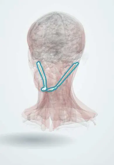
The digastric is a two-bellied muscle under the jaw, with a middle tendon looped to the hyoid bone; it opens the jaw and lifts the hyoid when swallowing.
Official name: Anterior belly of digastric muscle
Opens in the Durgical app, no sign-up needed.
What it is
A two-bellied muscle under the jaw, joined by a tendon looped to the hyoid.
What it does
Opens the jaw and lifts the hyoid when swallowing.
Key facts
- Origin
- Anterior belly: digastric fossa of the mandible; posterior belly: mastoid notch
- Insertion
- Intermediate tendon slung to the hyoid
- Action
- Depresses the mandible; elevates the hyoid
- Nerve
- Anterior belly: nerve to mylohyoid (V3); posterior belly: facial nerve (VII)
- Blood supply
- Submental artery, occipital artery
Relations
- Course
- The posterior belly runs down and forward from the mastoid notch of the temporal bone, passes through the split tendon of the stylohyoid, becomes the intermediate tendon held to the hyoid by a fibrous loop, and the anterior belly then runs forward to the inner chin
- Superficial
- Skin, platysma and the investing layer of the cervical fascia; the submandibular gland overlaps both bellies and the parotid gland touches the posterior belly
- Deep
- Posterior belly: internal and external carotid arteries, internal jugular vein, and the vagus, accessory and hypoglossal nerves; anterior belly: the mylohyoid
- Below
- The occipital artery runs back along the lower border of the posterior belly; the hypoglossal nerve emerges from under it
How to find it
The anterior belly is felt under the chin, just beside the midline, when the person opens the mouth against resistance. The posterior belly cannot be felt; in surgery it is found below the parotid gland, running from the mastoid process to the hyoid bone.
Examination
- Opening the mouth against a hand placed under the chin: both anterior bellies tighten beside the midline; a soft belly on one side, with a weak masseter, points to the mandibular nerve on that side
On imaging
On CT and MRI the anterior bellies lie under the mylohyoid on each side of the midline; the posterior belly runs from the mastoid notch toward the hyoid, superficial to the internal jugular vein and carotids. The medial edge of the anterior belly divides node level IA (submental) from IB (submandibular). A small, fatty anterior belly may mean denervation from a V3 lesion, not a mass.
Clinical notes
- Key landmark in submandibular gland excision, neck dissection and carotid surgery: dissection superficial to the posterior belly keeps the great vessels and cranial nerves IX–XII out of harm
- In parotidectomy, the facial nerve trunk is found just above the posterior belly where it attaches to the mastoid
- The anterior belly can be transferred to the lower lip to restore its movement after injury to the marginal mandibular branch of the facial nerve
Anatomical variants
- Accessory anterior bellies, sometimes crossing the midline under the chin, are the most commonly described variation and can look like a mass on imaging
Development
The anterior belly comes from the first pharyngeal arch (hence V3) and the posterior belly from the second (hence the facial nerve), which explains the double nerve supply.
Test yourself
Think of the answer, then tap the question to see it.
What is the nerve supply of each belly of the digastric, and why does it differ?
Anterior belly: nerve to mylohyoid (V3); posterior belly: facial nerve (VII). They come from the first and second pharyngeal arches.
Which triangle do the two bellies of the digastric outline with the lower border of the mandible?
The submandibular triangle; the two anterior bellies and the hyoid bone outline the submental triangle.
Why is the posterior belly of the digastric called the head and neck surgeon's «friend»?
The great vessels and the lower cranial nerves (IX–XII) lie deep to it, so dissection superficial to it is safe.
In parotidectomy, where is the facial nerve trunk found in relation to the digastric?
Just above the posterior belly of the digastric, where it attaches to the mastoid.
Educational information, not medical advice. For any symptom, see a health professional.
Its names
- Official Latin (TA2)
- venter anterior musculi digastrici TA2 2161
- Official Spanish (DeCS)
- Músculo digástrico
- English (TA2)
- Anterior belly of digastric muscle
- Also called
- venter mandibularis musculi digastrici
Related
Sources: Terminologia Anatomica (TA2); Anatomy, Head and Neck, Digastric Muscle; Anatomy, Head and Neck: Suprahyoid Muscle; 11.3 Axial Muscles of the Head, Neck, and Back (OpenStax (Rice University), 2022) · adapted, CC BY 4.0.
Spotted a mistake?We are always improving for the community. Tell us what is wrong and our team will review it.Report a mistake
Representative model, for educational purposes only.