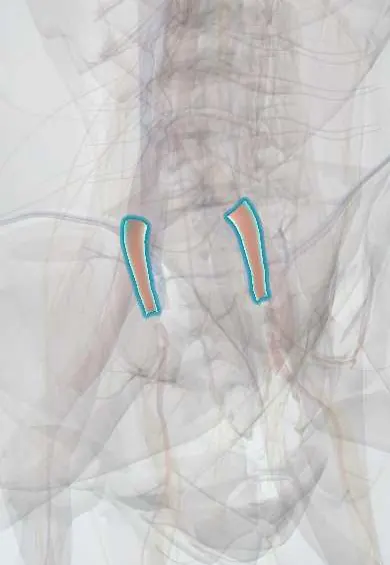
Internal iliac arteries !
The internal iliac arteries are the inner branches of the common iliac arteries; they supply the bladder, rectum, reproductive organs and buttocks.
Official name: Internal iliac artery
Opens in the Durgical app, no sign-up needed.
What it is
The inner branch of each common iliac artery, starting in front of the sacroiliac joint.
What it does
Supplies the bladder, rectum, reproductive organs, the gluteal muscles and the pelvic floor.
Key facts
- Origin
- Common iliac bifurcation in front of the sacroiliac joint (L5–S1 level)
- Branches
- Posterior: iliolumbar, lateral sacral, superior gluteal; anterior: umbilical, obturator, vesical, uterine, middle rectal, internal pudendal, inferior gluteal
- Supplies
- Pelvic viscera, perineum, gluteal region, medial thigh (obturator), lower spine (iliolumbar, lateral sacral)
Relations
- Course
- From the common iliac bifurcation in front of the sacroiliac joint it drops backward into the pelvis and, near the upper edge of the greater sciatic foramen, divides into anterior and posterior divisions
- In front
- Ureter and, in women, the ovary in the ovarian fossa
- Behind
- Internal iliac vein, the lumbosacral trunk and the sacroiliac joint
- Lateral
- External iliac vein and the obturator nerve
How to find it
It cannot be felt. At surgery it is found at the pelvic brim just behind the point where the ureter crosses; for embolization it is reached by a catheter, usually passed from a femoral artery.
On imaging
CT angiography shows it from the iliac bifurcation at the pelvic brim, where the ureter crosses in front, and in pelvic fractures it shows active bleeding as a contrast blush. Catheter angiography then confirms the bleeding branch and treats it by embolization. Ultrasound or CT finds an internal iliac aneurysm, often with an aortoiliac one, and CT checks for endoleak after a stent-graft.
Clinical notes
- In obstetric haemorrhage its anterior division can be tied or embolized; beside the cervix the uterine artery crosses above the ureter ("water under the bridge")
- Blocking both internal iliac arteries, for example to extend an aortic stent-graft, can cause buttock claudication and, less often, colon or spinal cord ischaemia
- Its posterior division (iliolumbar and lateral sacral arteries) feeds the lower lumbar and sacral nerve roots; sacral fractures and sacroiliac screws put the superior gluteal vessels at risk
- When it is tied (for example in pelvic or obstetric haemorrhage), the tie goes on its trunk a few centimetres from the origin, beyond the posterior division, to spare the gluteal arteries; the internal iliac vein lies right behind it and is the main danger
Anatomical variants
- The level of origin and the way it splits vary: most often the anterior division gives the inferior gluteal and internal pudendal arteries and the posterior division the superior gluteal; in about a fifth the two gluteal arteries share a trunk, and a true trifurcation is rare (series differ)
- The obturator artery often arises elsewhere, commonly from the inferior epigastric artery, crossing the pubic ramus as the "corona mortis", at risk in pelvic and hernia surgery
Development
In the fetus it is larger than the external iliac and continues as the umbilical artery to the placenta. After birth only its pelvic part stays open, as the internal iliac and the start of the superior vesical artery; the rest closes into the medial umbilical ligament, a guide to the artery in laparoscopic pelvic surgery.
Memory aid
Posterior division "PILS": Posterior = Iliolumbar, Lateral sacral, Superior gluteal; all the other branches come from the anterior division.
Related conditions
Test yourself
Think of the answer, then tap the question to see it.
Which branches come from the posterior division of the internal iliac artery?
Iliolumbar, lateral sacral and superior gluteal («PILS»); all other branches come from the anterior division.
Why is tying or blocking an internal iliac artery usually tolerated?
Collaterals: lumbar with iliolumbar, median with lateral sacral, and superior with middle rectal arteries.
Which branch of the internal iliac artery is most often injured in posterior pelvic ring fractures?
The superior gluteal artery, its largest branch, leaving above the piriformis through the greater sciatic notch.
Where does the uterine artery cross the ureter?
Beside the cervix, the uterine artery passes above the ureter («water under the bridge»).
What is the «corona mortis» related to the internal iliac artery?
An obturator artery arising from the inferior epigastric artery and crossing the pubic ramus, at risk in pelvic and hernia surgery.
Educational information, not medical advice. For any symptom, see a health professional.
Its names
- Official Latin (TA2)
- arteria iliaca interna TA2 4302
- Official Spanish (DeCS)
- Arteria ilíaca interna DeCS D007083
- English (TA2)
- Internal iliac artery
- Also called
- hypogastric artery · arteria hypogastrica · arteria ilica interna
Related
Sources: Terminologia Anatomica (TA2); DeCS; Anatomy, Abdomen and Pelvis: Internal Iliac Arteries; Anatomy of the Human Body (20th ed.): The Common Iliac Arteries (Lea & Febiger (Gray's Anatomy, 1918), 1918) · adapted, Public domain; 28.5 Adjustments of the Infant at Birth and Postnatal Stages (OpenStax (Rice University), 2022) · adapted, CC BY 4.0; Tratado de anatomía humana (9.ª ed.), tomo II, Angiología: aorta descendente, arterias ilíacas y arterias del miembro inferior.
Spotted a mistake?We are always improving for the community. Tell us what is wrong and our team will review it.Report a mistake
Representative model, for educational purposes only.