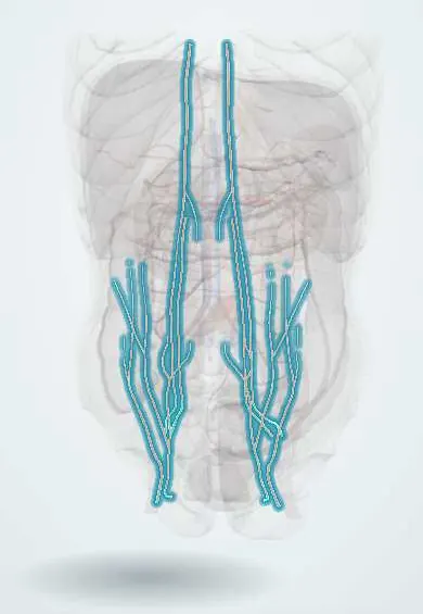
Cardiovascular system · Abdomen
Epigastric vessels !
The epigastric vessels are arteries and veins that run up behind the rectus abdominis; the lower ones are a key landmark for telling groin hernia types apart.
Official name: Inferior epigastric artery
Opens in the Durgical app, no sign-up needed.
What it is
Arteries and veins running up the inside of the abdominal wall behind the rectus muscles (inferior and superior epigastric) and in the fat (superficial epigastric).
What it does
Supply the abdominal wall. The inferior epigastric vessels are the landmark between direct (inside them) and indirect (outside them) hernias, and laparoscopic ports avoid them.
Key facts
- Origin
- Inferior: external iliac artery just above inguinal ligament; superior: internal thoracic artery (terminal branch); superficial: femoral artery
- Branches
- Inferior: cremasteric and pubic branches; superior and inferior anastomose inside rectus sheath; paired venae comitantes
- Supplies
- Rectus abdominis and anterior abdominal wall; inferior vessels raise lateral umbilical fold, medial to deep ring (indirect lateral, direct medial)
Relations
- Course
- Inferior: from the external iliac artery just above the inguinal ligament it curves up and medially, medial to the deep inguinal ring, pierces the transversalis fascia and enters the rectus sheath at the arcuate line, then climbs behind the muscle
- Lateral
- Deep inguinal ring, with the ductus deferens (or the round ligament) hooking around the artery as it enters the canal
- Medial
- Hesselbach triangle and the lateral border of the rectus abdominis
- Behind
- Transversalis fascia and parietal peritoneum, raised as the lateral umbilical fold
- In front
- Rectus abdominis, once inside the rectus sheath
How to find it
It cannot be felt. Its line runs from about the mid-inguinal point toward the umbilicus. At laparoscopy it is seen in the lateral umbilical fold, and Doppler ultrasound can map it before a drain or port is placed.
Examination
- Fothergill sign: a rectus sheath haematoma stays palpable and does not cross the midline when the abdominal muscles are tensed, unlike a mass inside the abdomen
On imaging
CT shows a rectus sheath haematoma as a spindle-shaped mass in the muscle, with active bleeding on the arterial phase. On CT or at laparoscopy, a hernia sac medial to the inferior epigastric vessels is direct; one lateral to them is indirect.
Clinical notes
- Paracentesis and drains are placed lateral to the rectus sheath, usually in the left lower quadrant, away from the inferior epigastric artery
- Deep inferior epigastric perforator (DIEP) flap: lower abdominal skin and fat carried on perforators of this artery to rebuild the breast
- The superficial epigastric veins dilate as a collateral in inferior vena cava obstruction, with flow upward from below the umbilicus
Anatomical variants
- An obturator artery arising from the inferior epigastric artery and crossing near the femoral ring (corona mortis) is at risk in femoral hernia repair and pelvic fracture surgery; a venous link between the inferior epigastric (or external iliac) and obturator veins over the pubic ramus is even more frequent and bleeds just as readily
Memory aid
MDs don't LIe: Medial to the vessels is Direct, Lateral is Indirect.
Related conditions
Test yourself
Think of the answer, then tap the question to see it.
Where does the inferior epigastric artery arise and where does it enter the rectus sheath?
From the external iliac artery just above the inguinal ligament; it enters the rectus sheath at the arcuate line and climbs behind the rectus abdominis.
Which peritoneal fold do the inferior epigastric vessels raise?
The lateral umbilical fold, just medial to the deep inguinal ring.
Why are paracentesis needles and drains placed lateral to the rectus sheath?
To avoid the inferior epigastric artery, which runs behind the rectus abdominis; the left lower quadrant is the usual site.
Why do the epigastric arteries enlarge in coarctation of the aorta?
The superior (from the internal thoracic) and inferior epigastric arteries link the subclavian and external iliac arteries, forming a bypass around the narrowing.
Educational information, not medical advice. For any symptom, see a health professional.
Its names
- Official Latin (TA2)
- arteria epigastrica inferior; arteria epigastrica superior; arteria epigastrica superficialis TA2 4358
- Official Spanish (DeCS)
- Vasos epigástricos DeCS D019074
- English (TA2)
- Inferior epigastric artery
- Also called
- arteria epigastrica caudalis
Related
Sources: Terminologia Anatomica (TA2); DeCS; Anatomy, Abdomen and Pelvis: Epigastric Artery; Tratado de anatomía humana (9.ª ed.), tomo II, Libro IV Angiología, sección 4: Venas (sistemas de las venas cavas superior e inferior, vena porta, venas del raquis).
Spotted a mistake?We are always improving for the community. Tell us what is wrong and our team will review it.Report a mistake
Representative model, for educational purposes only.