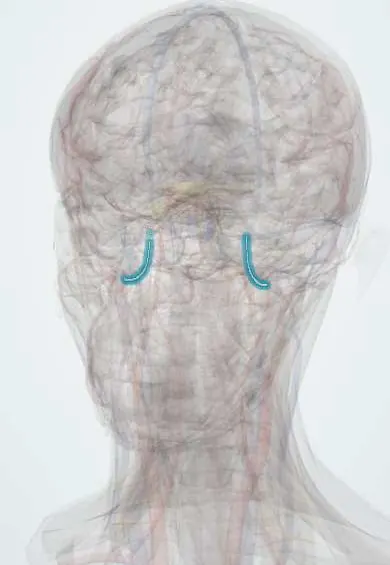
Epidural haematoma !
An epidural haematoma is bleeding between the skull and the brain's outer covering, usually after a skull fracture; it can grow fast and is an emergency.
Updated 2026-10-29
Structures involved
Tap one to see what it is and open it in 3D.
- Middle meningeal artery
- Dura mater
- Pterion
- Temporal bones
- Parietal bones
- Foramen spinosum
- Temporal lobe
- Oculomotor nerve (III)
- Midbrain
In the clinical exam
In physiology
Why it happens
- Hemorrhage: petechiae, purpura, ecchymosis, hematoma, and what blood loss does
- Mechanical and electrical injury: abrasion, contusion, laceration, wounds
What it is
A collection of blood between the inner table of the skull and the dura mater, usually after a skull fracture tears the middle meningeal artery. It is a neurosurgical emergency because it can expand within hours.
Who gets it
Mostly young adults and adolescents after road traffic accidents, falls and assaults. It is uncommon in older adults, in whom the dura is more firmly attached to the skull. Motorcycle accidents are an important cause in Colombia.
Causes
- Temporal or temporoparietal skull fracture tearing the middle meningeal artery
- Venous bleeding from dural sinuses or diploic veins (less common, slower; posterior fossa)
- Road traffic accidents, falls, assaults, sports injuries
Risk factors
- Young age
- Motorcycle riding without a helmet
- High-energy head trauma
- Alcohol use at the time of injury
- Anticoagulant therapy (worsens bleeding)
Pathophysiology
The middle meningeal artery runs in a groove on the inner skull near the pterion, where the bone is thin. Arterial blood strips the dura off the bone, forming a lens-shaped collection limited by the sutures. As it grows, intracranial pressure rises and the temporal lobe can herniate under the tentorium, compressing the oculomotor nerve and midbrain.
Symptoms and signs
- Head injury, often with brief loss of consciousness
- Lucid interval: the patient wakes and seems well, then deteriorates (in only a minority of cases)
- Worsening headache, vomiting, drowsiness
- Dilated pupil on the side of the clot (oculomotor nerve compression)
- Contralateral weakness
- Bradycardia, hypertension and irregular breathing late (Cushing response)
Diagnosis
- Clinical
- Trauma history plus neurological examination with serial Glasgow Coma Scale scoring and pupil checks. A lucid interval is classic but its absence does not exclude the diagnosis.
- Tests
- Coagulation studies, full blood count, blood group and crossmatch before surgery; blood alcohol and toxicology when consciousness is impaired.
- Imaging
- Non-contrast head CT is the test of choice: a biconvex (lens-shaped) hyperdense collection that does not cross suture lines, often with an overlying fracture. Mixed density suggests active bleeding.
Differential diagnosis
- Acute subdural haematoma
- Cerebral contusion
- Traumatic subarachnoid haemorrhage
- Intracerebral haemorrhage
- Diffuse axonal injury
Goal of treatment
Remove the clot and stop the bleeding before the brain herniates, while protecting airway, breathing, circulation and cerebral perfusion.
Treatment
- Acute
- Trauma resuscitation (airway with cervical spine protection, breathing, circulation), head elevation and avoidance of hypoxia and hypotension; reversal of anticoagulation.
- Surgical
- Urgent craniotomy and evacuation for large or symptomatic haematomas, especially with a falling consciousness level or pupil changes.
- Acute
- Temporary measures for raised intracranial pressure, such as hyperosmolar therapy, while surgery is arranged.
- Follow-up
- Small haematomas in alert patients without mass effect may be observed in a neurosurgical unit with serial neurological checks and repeat CT.
Complications
- Transtentorial (uncal) herniation
- Death
- Seizures
- Persistent neurological deficits
- Infection after surgery
Prognosis
With early recognition and surgery, outcome is often good because the brain beneath may be little injured. Prognosis worsens with a low Glasgow score, abnormal pupils and delay to surgery.
Prevention
- Helmet use on motorcycles and bicycles
- Seat belts
- Fall prevention
- Road safety measures
Test yourself
Think of the answer, then tap the question to see it.
Which vessel is classically torn in an epidural haematoma?
The middle meningeal artery, often from a fracture of the thin bone at the pterion.
What is the CT shape of an epidural haematoma and why?
Biconvex (lens-shaped): the dura is firmly attached at the sutures, so the blood cannot spread past them.
What is the lucid interval?
A period of near-normal consciousness after the initial injury, followed by deterioration as the haematoma enlarges.
Why does an expanding temporal epidural haematoma dilate the ipsilateral pupil?
Uncal herniation compresses the oculomotor nerve, whose outer parasympathetic fibres constrict the pupil.
Related conditions
Educational information, not medical advice. For any symptom, see a health professional.
Sources: Epidural Hematoma (StatPearls (NCBI Bookshelf), 2025).
Spotted a mistake?We are always improving for the community. Tell us what is wrong and our team will review it.Report a mistake
Representative model, for educational purposes only.