Reproductive system · Pelvis · Female
Rectouterine pouch !
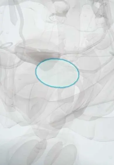
The rectouterine pouch (of Douglas) is the space between the uterus and the rectum, the lowest point of the abdomen; fluid or blood collects here first.
Opens in the Durgical app, no sign-up needed.
What it is
The pocket of peritoneum between the back of the uterus and the rectum (pouch of Douglas), the lowest point of the abdominal cavity when standing.
What it does
Fluid or blood collects here first, so it is checked on ultrasound; a common site of endometriosis.
Key facts
- Where
- Peritoneal recess between the posterior uterus/posterior fornix and the rectum, bounded laterally by the uterosacral folds
- Function
- Lowest point of the peritoneal cavity (upright): fluid, blood or pus collects here; reached through the posterior fornix; common site of endometriosis
Relations
- In front
- Back of the uterus and supravaginal cervix, and the peritoneum over the posterior vaginal fornix
- Behind
- Front of the middle third of the rectum
- Lateral
- Uterosacral folds raised by the uterosacral ligament; the ovaries and uterine tubes may drop into it
- Below
- Rectovaginal septum, between the vagina and the rectum
- Contents
- Normally loops of ileum or sigmoid colon; in disease, fluid, blood, pus, endometriosis or tumour deposits
How to find it
Transvaginal ultrasound shows it in the sagittal plane behind the uterus and above the posterior fornix. On rectal or vaginal examination its contents are felt through the front wall of the rectum or the posterior fornix.
Examination
- Douglas cry: sharp pain when the pouch is pressed through the posterior fornix or the rectum, suggesting blood or pus irritating the pelvic peritoneum
On imaging
On sagittal transvaginal ultrasound it lies behind the uterus. A small amount of clear fluid is normal; echogenic fluid or clot with a positive pregnancy test and no intrauterine pregnancy suggests a ruptured ectopic. In trauma, the pelvic FAST view checks it through a full bladder. Transvaginal ultrasound and MRI show deep endometriosis obliterating it.
Common problems
- Weakened supports allow prolapse.
- Endometriosis often grows on the uterosacral ligaments.
How it is diagnosed
Examination and ultrasound; seen directly in laparoscopy.
Treatment
- They are divided step by step in a hysterectomy.
Clinical notes
- Culdocentesis, a needle through the posterior fornix into the pouch, was used to find blood (for example from a ruptured ectopic pregnancy); ultrasound has largely replaced it
- Pelvic abscesses after appendicitis, pelvic inflammatory disease or surgery collect here and may be drained through the vagina or the rectum
- The pelvic view of the FAST scan looks for free fluid here in women and in the rectovesical pouch in men
- Peritoneal metastases deposited here form a shelf felt on rectal or vaginal examination (Blumer shelf)
- A deep pouch can herniate down into the rectovaginal septum as an enterocele, a form of posterior compartment prolapse
Related conditions
Test yourself
Think of the answer, then tap the question to see it.
What is the lowest point of the peritoneal cavity in an upright woman?
The rectouterine pouch (pouch of Douglas), between the back of the uterus and the rectum.
Why can the rectouterine pouch be reached through the vagina?
Its floor lies against the posterior vaginal fornix: today by transvaginal ultrasound and posterior colpotomy, formerly by culdocentesis.
What is the Douglas cry?
Sharp pain when the rectouterine pouch is pressed through the posterior fornix or rectum, suggesting blood or pus irritating the pelvic peritoneum.
What is a Blumer shelf?
Peritoneal metastases in the rectouterine pouch forming a shelf felt on rectal or vaginal examination.
Educational information, not medical advice. For any symptom, see a health professional.
Its names
- Official Latin (TA2)
- excavatio rectouterina TA2 3726
- Official Spanish (DeCS)
- Excavación rectouterina
- English (TA2)
- Rectouterine pouch
- Also called
- cavum Douglasi · pouch of Douglas
Related
Sources: Terminologia Anatomica (TA2); Anatomy, Abdomen and Pelvis: Female Pelvic Cavity; Anatomy, Abdomen and Pelvis, Peritoneum.
Spotted a mistake?We are always improving for the community. Tell us what is wrong and our team will review it.Report a mistake
Representative model, for educational purposes only.