Conditions · Nephrology and urology
Renal papillary necrosis !
Death of the tips of the kidney pyramids (papillae). Pieces can slough into the urine and cause blood in the urine, colicky pain or blockage.
Updated 2026-11-01
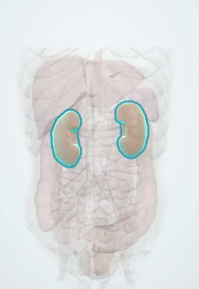
Structures involved
Tap one to see what it is and open it in 3D.
Why it happens
- Patterns of necrosis: coagulative, liquefactive, caseous, fat, fibrinoid, gangrenous
- Injury by therapeutic drugs: the classic examples
- The countercurrent mechanism, urea recycling and urine concentration
- Kidney: cortex, medulla and the nephron
What it is
Ischaemic necrosis of the renal papillae, the innermost tips of the medullary pyramids that drain into the calyces. The dead tissue may slough and pass down the ureter, causing haematuria, colic and obstruction.
Who gets it
Uncommon and often found at autopsy or on imaging. It mainly affects adults with diabetes, people with long-term heavy analgesic use and people with sickle cell disease or trait, a relevant group in Afro-Colombian communities of the Pacific and Caribbean coasts.
Causes
- Pyelonephritis, especially in diabetes or with obstruction
- Long-term heavy use of analgesics (classically combinations with phenacetin; also NSAIDs)
- Sickle cell disease or sickle cell trait
- Diabetes mellitus
- Urinary tract obstruction
- Kidney transplant rejection, severe dehydration or shock, and renal tuberculosis
Risk factors
- Diabetes, especially long-standing
- Frequent or prolonged use of over-the-counter pain relievers
- Sickle haemoglobin (disease or trait)
- Urinary infection with obstruction or stones
- Dehydration and older age
Pathophysiology
The papillae receive their blood from the long, thin vasa recta and live in a hypoxic, hypertonic environment, so they are the most vulnerable part of the kidney to ischaemia. Sickled cells occlude the vasa recta in the hypoxic medulla, diabetes damages small vessels, NSAIDs block the prostaglandins that dilate medullary vessels, and analgesic metabolites concentrate in the papillae. Infection and obstruction raise pressure and add inflammatory injury. The tip dies, may separate and either stays in the calyx or travels down the ureter.
Classification
- Medullary (partial) form: necrosis within the papilla with the tip remaining in place
- Papillary (total) form: the whole papilla sloughs into the calyx
- Mnemonic of causes: POSTCARDS (pyelonephritis, obstruction, sickle cell, tuberculosis, cirrhosis, analgesics, renal transplant rejection, diabetes, systemic vasculitis)
Symptoms and signs
- Gross haematuria
- Flank pain or ureteric colic
- Passage of tissue fragments in the urine
- Fever and chills when infection is present
- Reduced urine output if both kidneys are obstructed
- May be silent and show only as microscopic haematuria or reduced concentrating ability
Diagnosis
- Clinical
- Suspect it with haematuria and flank pain in a person with diabetes, sickle cell disease, analgesic overuse or pyelonephritis.
- Tests
- Urinalysis (blood, white cells, sometimes necrotic tissue), urine culture, creatinine and electrolytes; pathology of passed fragments confirms necrotic papilla. Haemoglobin electrophoresis if sickle status is unknown.
- Imaging
- CT urography is the test of choice: contrast pooling in cavities of the papillae, "ring sign" around a sloughed papilla and filling defects. Ultrasound can show obstruction and hydronephrosis.
Differential diagnosis
- Kidney stones
- Acute pyelonephritis without necrosis
- Urothelial carcinoma of the renal pelvis
- Blood clot in the collecting system
- Renal tuberculosis
Goal of treatment
Remove the cause, treat infection, relieve obstruction and preserve kidney function.
Treatment
- Medical
- Stop analgesics such as NSAIDs and phenacetin-containing combinations; optimise glucose control and manage sickle cell disease.
- Medical
- Antibiotics guided by urine culture when infection is present; hydration and pain control.
- Procedure
- Relief of obstruction from a sloughed papilla with a ureteral stent, ureteroscopy or percutaneous nephrostomy.
- Acute
- Urgent care for urosepsis or acute kidney injury with obstruction: fluids, intravenous antibiotics and emergency drainage.
- Follow-up
- Follow-up of kidney function, urinalysis and blood pressure; review all over-the-counter medicines.
Complications
- Ureteral obstruction and hydronephrosis
- Urosepsis
- Acute kidney injury and chronic kidney disease
- Urothelial carcinoma in analgesic nephropathy
Prognosis
It depends on the cause and on how many papillae are affected. Removing the cause (stopping analgesics, treating infection) can stop further damage, but necrosis is irreversible and severe bilateral disease can lead to chronic kidney disease.
Prevention
- Avoiding long-term, unsupervised use of pain relievers
- Good diabetes control and prompt treatment of urinary infections
- Adequate hydration in people with sickle cell disease or trait
Test yourself
Think of the answer, then tap the question to see it.
Why are the renal papillae vulnerable to ischaemia?
They are supplied by the long vasa recta in a hypoxic, hypertonic medulla, at the end of the blood supply.
What are four classic causes of renal papillary necrosis?
Diabetes, analgesic overuse, sickle cell disease or trait and acute pyelonephritis (also obstruction, tuberculosis and transplant rejection).
Typical presentation?
Gross haematuria and flank pain or colic, sometimes with passage of tissue in the urine.
Best imaging test?
CT urography, which may show a ring sign or filling defects.
Related conditions
- Acute pyelonephritis
- Sickle cell disease
- Type 2 diabetes mellitus
- Type 1 diabetes mellitus
- Acute kidney injury
- Chronic kidney disease
- Kidney stones (urolithiasis)
- Hydronephrosis and obstructive uropathy
- Kidney transplant rejection
- Acute interstitial nephritis
- Sepsis and septic shock
Educational information, not medical advice. For any symptom, see a health professional.
Sources: Renal papillary necrosis: MedlinePlus Medical Encyclopedia (MedlinePlus (U.S. National Library of Medicine), 2024).
Spotted a mistake?We are always improving for the community. Tell us what is wrong and our team will review it.Report a mistake
Representative model, for educational purposes only.