Conditions · Nephrology and urology
Hydronephrosis and obstructive uropathy !
Hydronephrosis is swelling of the kidney's collecting system when urine cannot drain, usually from a blockage such as a stone or an enlarged prostate.
Updated 2026-11-01
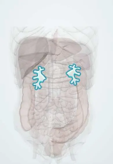
Structures involved
Tap one to see what it is and open it in 3D.
In the clinical exam
Why it happens
- Atrophy
- Glomerular filtration: the barrier and its determinants
- Physiology of acute and chronic kidney failure
- Ureter, bladder and urethra: urothelium
- Kidney: cortex, medulla and the nephron
What it is
Hydronephrosis is dilation of the renal pelvis and calyces; when the ureter is also dilated it is called hydroureteronephrosis. Obstructive uropathy is the blockage of urine flow anywhere from the kidney to the urethra, and obstructive nephropathy is the kidney damage it causes. Obstruction can be one-sided or both-sided, acute or chronic, partial or complete.
Who gets it
Found at all ages. In infants it is often detected on prenatal ultrasound (pelvi-ureteric junction obstruction, vesicoureteral reflux, posterior urethral valves in boys). In young adults stones dominate; in older men, prostate enlargement; in women, pregnancy and pelvic tumours such as cervical cancer, which remains a frequent cancer in Colombia and Latin America and a classic cause of bilateral ureteric obstruction.
Causes
- Kidney or ureteric stones
- Benign prostatic hyperplasia, prostate cancer or urethral stricture (bladder outlet obstruction)
- Pelvic tumours: cervical, bladder, colorectal or ovarian cancer; retroperitoneal fibrosis or lymph nodes
- Congenital: pelvi-ureteric junction obstruction, posterior urethral valves, vesicoureteral reflux
- Neurogenic bladder (spinal cord injury, diabetes, multiple sclerosis)
- Pregnancy (physiological dilation, more on the right) and ureteric injury after pelvic surgery
- Blood clots, sloughed papillae or ureteric strictures
Risk factors
- Previous kidney stones
- Male sex and older age (prostate disease)
- Pelvic cancer, radiotherapy or surgery
- Neurological disease affecting bladder emptying
- Pregnancy
- Single functioning kidney (any obstruction is critical)
Pathophysiology
Blocked outflow raises pressure in the ureter and pelvis, which is transmitted back to the tubules and lowers glomerular filtration. Early on, renal blood flow rises, then falls as vasoconstriction follows. Prolonged pressure causes tubular atrophy, interstitial inflammation and fibrosis, so function lost after weeks may not return. Stagnant urine also favours infection and stone formation. After relief of a bilateral obstruction, the damaged tubules may lose large amounts of water and salt (post-obstructive diuresis).
Classification
- Unilateral versus bilateral (or obstruction of a single kidney)
- Acute versus chronic; partial versus complete
- Upper tract (kidney, ureter) versus lower tract (bladder outlet, urethra)
- Ultrasound grading of dilation (mild, moderate, severe) and, in infants, the SFU or UTD systems
Symptoms and signs
- Acute: colicky flank pain radiating to the groin, nausea and vomiting (stones)
- Chronic: often silent, or a dull flank ache
- Reduced or absent urine output (bilateral or bladder outlet obstruction)
- Weak stream, hesitancy, frequency and incomplete emptying (prostate)
- Palpable bladder or flank mass
- Haematuria, fever and dysuria when infection is present
- Hypertension or signs of kidney failure in long-standing obstruction
Diagnosis
- Clinical
- Pain pattern, urinary symptoms, palpable bladder, digital rectal examination in men and pelvic examination in women; review of past stones, cancers, surgery and neurological disease.
- Tests
- Creatinine, urea and electrolytes (potassium); complete blood count; urinalysis and urine culture; post-void residual volume by bladder scan; PSA where relevant.
- Imaging
- Kidney and bladder ultrasound is the first test (no radiation, safe in pregnancy and kidney failure). Non-contrast CT identifies stones and the level of blockage; contrast CT or MRI urography for tumours; diuretic renography (MAG3) measures split function and true obstruction; voiding cystourethrogram in children.
Differential diagnosis
- Physiological dilation in pregnancy
- Vesicoureteral reflux without obstruction
- Extrarenal pelvis (a normal variant)
- Parapelvic kidney cysts
- Polycystic kidney disease
Goal of treatment
Relieve the obstruction quickly, treat infection, protect kidney function and correct the cause so it does not return.
Treatment
- Acute
- Infected obstruction is an emergency: intravenous antibiotics plus urgent drainage. Pain relief with NSAIDs or opioids and antiemetics for colic.
- Procedure
- Drainage: urinary catheter for bladder outlet obstruction; ureteric stent or percutaneous nephrostomy for upper tract obstruction.
- Surgical
- Definitive treatment of the cause: ureteroscopy, lithotripsy or percutaneous removal of stones; prostate surgery; pyeloplasty for pelvi-ureteric junction obstruction; oncological treatment of pelvic tumours.
- Medical
- Medical expulsive therapy with alpha-blockers for small distal ureteric stones; alpha-blockers and 5-alpha-reductase inhibitors for prostate enlargement.
- Follow-up
- Nursing care: urine output and fluid balance, care of catheters and nephrostomy tubes, signs of infection, and electrolytes during post-obstructive diuresis.
Complications
- Acute kidney injury and chronic kidney disease
- Pyelonephritis, pyonephrosis and urosepsis
- Post-obstructive diuresis with dehydration and electrolyte loss
- Hyperkalaemia and metabolic acidosis
- Hypertension
- Stone formation
Prognosis
Function usually recovers if obstruction is relieved within days to a few weeks. Long-standing complete obstruction can cause permanent loss of the affected kidney. Bilateral obstruction from advanced pelvic cancer carries a poor outlook driven by the cancer.
Prevention
- Prevention of stone recurrence (fluids, diet, treating metabolic causes)
- Early evaluation of urinary symptoms in men
- Follow-up of prenatal hydronephrosis in infants
- Cervical cancer screening and HPV vaccination
Test yourself
Think of the answer, then tap the question to see it.
What is the first-line imaging test for suspected hydronephrosis?
Kidney and bladder ultrasound.
Why is an obstructed, infected kidney a urological emergency?
It can rapidly cause urosepsis; it needs intravenous antibiotics plus urgent drainage by stent or nephrostomy.
What is the most common cause of bilateral hydronephrosis in an older man?
Bladder outlet obstruction from benign prostatic hyperplasia.
What should nurses watch for after relieving a bilateral obstruction?
Post-obstructive diuresis: very high urine output with dehydration and loss of sodium and potassium.
Which congenital cause of hydronephrosis affects only boys?
Posterior urethral valves.
Related conditions
- Kidney stones (urolithiasis)
- Benign prostatic hyperplasia
- Acute urinary retention
- Acute pyelonephritis
- Acute kidney injury
- Chronic kidney disease
- Prostate cancer
- Cervical cancer
- Bladder cancer
- Neurogenic bladder
- Posterior urethral valves
- Congenital kidney anomalies (horseshoe kidney, renal agenesis)
- Urinary tract infection (acute cystitis)
Educational information, not medical advice. For any symptom, see a health professional.
Sources: Obstructive uropathy: MedlinePlus Medical Encyclopedia (MedlinePlus (U.S. National Library of Medicine), 2026); Bilateral hydronephrosis: MedlinePlus Medical Encyclopedia (MedlinePlus (U.S. National Library of Medicine), 2025).
Spotted a mistake?We are always improving for the community. Tell us what is wrong and our team will review it.Report a mistake
Representative model, for educational purposes only.