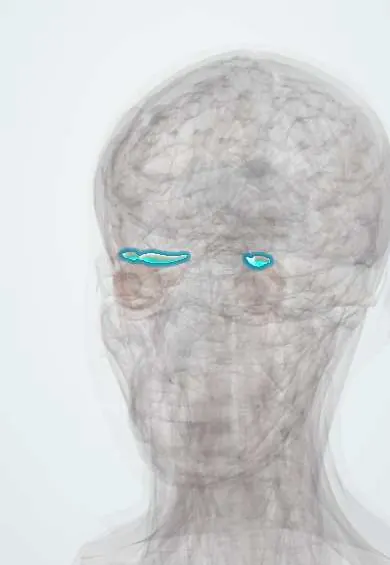
Superior rectus !
The superior rectus is one of the six muscles that move the eyeball, on top of the eye inside the eye socket; it turns the eye up and slightly in.
Official name: Superior rectus muscle
Opens in the Durgical app, no sign-up needed.
What it is
One of the six muscles that move the eyeball, inside the eye socket.
What it does
Turns the eye up (and slightly in).
Key facts
- Origin
- Common tendinous ring (annulus of Zinn)
- Insertion
- Sclera above, ~7.7 mm behind the limbus
- Action
- Elevation (best in abduction); intorsion, adduction
- Nerve
- Oculomotor nerve (III), superior division
- Blood supply
- Ophthalmic artery (muscular branches)
Relations
- Above
- Levator palpebrae superioris, which shares its sheath; above that, the frontal nerve and the orbital roof
- Below
- Globe and, further back, the optic nerve; the nasociliary nerve and ophthalmic artery cross over the optic nerve beneath it
- Medial
- Superior oblique, whose tendon passes under the superior rectus to insert behind it
- Lateral
- Lateral rectus and, at the front, the lacrimal gland
How to find it
Its tendon meets the sclera about 7.7 mm behind the upper limbus, farther back than any other rectus; at surgery it is hooked through the upper conjunctiva and the fascial sheath. Seen from above in dissection, it lies straight under the levator palpebrae superioris, joined to it by fascia, and the lacrimal gland rests on its lateral part.
Examination
- H-pattern (six cardinal gazes): the person follows a target out and then up; weak elevation in abduction isolates the superior rectus
- Diplopia pattern: vertical double vision greatest looking up and out toward the side of the weak muscle
On imaging
Coronal CT or MRI of the orbit shows it best: the four recti ring the optic nerve, and the superior rectus and levator lie together at the top as the superior muscle complex. Thyroid eye disease swells the muscle bellies and spares the tendons, usually inferior and medial rectus first, superior later. MRI may show a thin, atrophic muscle in a long-standing third nerve palsy.
Common problems
- Sixth-nerve palsy: the eye cannot turn out, causing side-by-side double vision.
- Strabismus (squint) when the eye muscles are unbalanced.
How it is diagnosed
Eye movement and alignment tests; scans if a nerve palsy is new.
Treatment
- Prisms or patching; botulinum toxin; eye muscle surgery to rebalance the muscles.
Clinical notes
- Complete oculomotor (III) palsy: eye down and out (unopposed lateral rectus and superior oblique), ptosis and, if the nerve is compressed, a dilated pupil
- A lesion of the superior division of the oculomotor nerve, near the orbital apex, weakens only the superior rectus and levator: ptosis with poor upgaze
- Thyroid eye disease can enlarge it (less often than the inferior and medial recti); a tight superior rectus limits downgaze
- Monocular elevation deficiency (double elevator palsy): a congenital weakness of upgaze in one eye, often with ptosis
- Its sheath is linked to the levator, so a large recession or resection of the superior rectus can shift the height of the upper lid
Development
Comes from the single mass of head mesoderm, beside the optic stalk, that forms every muscle of the oculomotor nerve; the levator palpebrae later separates from it.
Memory aid
"RAD SIN": the superior and inferior Recti ADduct; the Superior muscles (rectus and oblique) INtort.
Test yourself
Think of the answer, then tap the question to see it.
What are the actions of the superior rectus in primary gaze, and when is it a pure elevator?
Elevation, intorsion and adduction; with the eye abducted it is a pure elevator.
In which gaze direction is the superior rectus tested in isolation?
Looking out, then up: weak elevation in abduction isolates the superior rectus.
Which nerve supplies the superior rectus, and what is unusual about its nucleus?
The superior division of the oculomotor nerve (III); its fibres cross, so a nuclear lesion weakens the opposite superior rectus.
What does a lesion of the superior division of the oculomotor nerve produce?
Weakness of only the superior rectus and levator: ptosis with poor upgaze.
Educational information, not medical advice. For any symptom, see a health professional.
Its names
- Official Latin (TA2)
- musculus rectus superior TA2 2042
- Official Spanish (DeCS)
- Músculo recto superior DeCS D009801
- English (TA2)
- Superior rectus muscle
- Also called
- musculus rectus superior bulbi
Related
Sources: Terminologia Anatomica (TA2); DeCS; Anatomy, Head and Neck: Eye Superior Rectus Muscle; Anatomy, Head and Neck, Eye Extraocular Muscles; Anatomy of the Human Body (20th ed.): The Accessory Organs of the Eye (Lea & Febiger (Gray's Anatomy, 1918), 1918) · adapted, Public domain; Anatomy of the Human Body (20th ed.): Development of the Muscles (Lea & Febiger (Gray's Anatomy, 1918), 1918) · adapted, Public domain.
Spotted a mistake?We are always improving for the community. Tell us what is wrong and our team will review it.Report a mistake
Representative model, for educational purposes only.