Reproductive system · Pelvis · Male
Seminal vesicles !
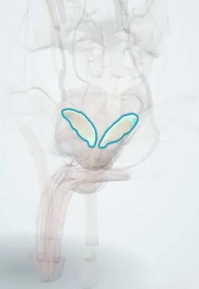
The seminal vesicles are two coiled glands behind the bladder; they make most of the fluid of semen, rich in fructose that feeds the sperm.
Official name: Seminal gland
Opens in the Durgical app, no sign-up needed.
What it is
Two coiled, sac-like glands, about 5 cm long, behind the bladder and above the prostate.
What it does
Make most of the fluid of semen (rich in fructose, an energy source for sperm); each joins a ductus deferens to form an ejaculatory duct.
Key facts
- Where
- Paired coiled sacs behind the bladder base and above the prostate, lateral to the ampullae of the ductus deferens; in front of the rectum
- Blood supply
- Inferior vesical and middle rectal arteries; artery to the ductus deferens
- Nerve
- Pelvic plexus: sympathetic fibres (T10–L2) contract its smooth muscle at emission
- Function
- Alkaline, fructose-rich fluid that makes up most of the semen volume (about 60–70%); its duct joins the ductus deferens to form the ejaculatory duct; invasion by prostate cancer raises the stage
Relations
- In front
- Back (base) of the urinary bladder
- Behind
- Rectum, separated by the rectoprostatic (Denonvilliers) fascia
- Medial
- Ampullae of the ductus deferens, which lie between the two vesicles
- Below
- Base of the prostate, where each duct joins a ductus deferens to form an ejaculatory duct
- Above
- Ureters, which reach the bladder near their upper tips, and the rectovesical pouch
How to find it
Usually out of reach on digital rectal examination; when enlarged, firm or invaded by tumour they may be felt above the prostate.
On imaging
Transrectal ultrasound shows them as paired, symmetric structures above the prostate, like a bow tie. On T2-weighted MRI they are bright and convoluted; loss of that bright signal, a mass or asymmetry suggests invasion by prostate cancer. Dilated vesicles can point to ejaculatory duct obstruction.
Common problems
- Rarely diseased on their own; they can be inflamed together with the prostate.
- Prostate cancer can spread into them, which changes its stage.
How it is diagnosed
MRI of the prostate; transrectal ultrasound.
Treatment
- Removed together with the prostate in a radical prostatectomy.
Clinical notes
- Blood in the semen (haematospermia) usually comes from the seminal vesicles or prostate and in young men is mostly benign
- Their tips lie close to the pelvic plexus, so their dissection in prostatectomy can harm the nerves of erection
Anatomical variants
- Agenesis is rare and often comes with other mesonephric anomalies on the same side, such as renal agenesis; a seminal vesicle cyst with an absent kidney on that side is Zinner syndrome
Development
Bud from the lower end of the mesonephric (Wolffian) duct, which also gives the ureteric bud; this explains their link with kidney and ureter anomalies on the same side.
Related conditions
Test yourself
Think of the answer, then tap the question to see it.
Do the seminal vesicles store sperm?
No; they secrete alkaline, fructose-rich fluid making up most of the semen volume, including proteins that make semen clot.
What stage does seminal vesicle invasion by prostate cancer represent?
T3b; it worsens the outlook, and the vesicles are removed with the prostate in radical prostatectomy.
What is Zinner syndrome?
A seminal vesicle cyst with an absent kidney on the same side, reflecting their shared mesonephric (Wolffian) duct origin.
Where does haematospermia usually come from, and what is its usual nature in young men?
From the seminal vesicles or prostate; in young men it is mostly benign.
Educational information, not medical advice. For any symptom, see a health professional.
Its names
- Official Latin (TA2)
- glandula vesiculosa
- Official Spanish (DeCS)
- Glándula seminal
- English (TA2)
- Seminal gland
- Also called
- seminal vesicle · seminal glands
Related
- Urinary bladder
- Rectovesical pouch
- Ejaculatory ducts
- Ductus deferens
- Pelvic plexus and cavernous nerves
- Internal iliac arteries
- Inferior vesical and prostatic arteries
Sources: Anatomy, Abdomen and Pelvis, Seminal Vesicle.
Spotted a mistake?We are always improving for the community. Tell us what is wrong and our team will review it.Report a mistake
Representative model, for educational purposes only.