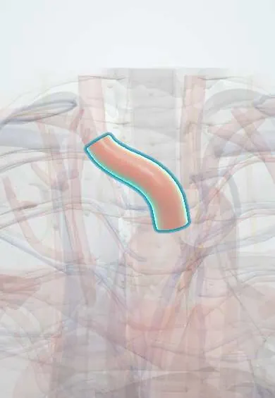
Brachiocephalic trunk !
The brachiocephalic trunk is the first branch of the aortic arch, on the right only; it splits into arteries for the right side of the head and right arm.
Opens in the Durgical app, no sign-up needed.
What it is
The first and largest branch of the aortic arch, on the right.
What it does
Splits into the right common carotid artery (to the head) and the right subclavian artery (to the arm).
Key facts
- Origin
- First and largest branch of aortic arch, behind manubrium
- Branches
- Right common carotid and right subclavian arteries, dividing behind right sternoclavicular joint; occasionally thyroid ima
- Supplies
- Right side of head and neck; right upper limb
Relations
- Course
- From the arch behind the manubrium it ascends upward, backward and to the right, first in front of and then to the right of the trachea, ending behind the right sternoclavicular joint
- In front
- Left brachiocephalic vein crossing it, the thymic remnant, the origins of the sternohyoid and sternothyroid muscles, and the manubrium
- Behind
- Trachea below; the right pleura higher up
- Lateral
- On its right: the right brachiocephalic vein, the upper superior vena cava and the right pleura
- Medial
- On its left: the left common carotid artery and the thymic remnant
How to find it
Not palpable in most adults. Its division projects onto the right sternoclavicular joint; in children, or when the trunk is long or tortuous, it can rise above the jugular notch and be felt pulsating there.
On imaging
CT or MR angiography shows it as the first, largest arch branch, rising to the right in front of the trachea to divide behind the right sternoclavicular joint. Look for a shared origin with the left common carotid (bovine arch), stenosis, aneurysm, or a high trunk in the jugular notch before tracheostomy. Neck Doppler shows reversed right vertebral flow in innominate steal.
Clinical notes
- Tracheo-innominate fistula: a tracheostomy tube tip or cuff erodes into the artery; a small sentinel bleed may come before a massive one
- A high-riding trunk in the jugular notch is at risk during tracheostomy and neck surgery and can be mistaken for an aneurysm
- Stenosis of the trunk can reverse flow in the right vertebral artery (innominate steal), with arm effort symptoms and dizziness
- In infants the trunk can compress the front of the trachea (innominate artery compression syndrome), a cause of stridor
Anatomical variants
- Shares a common origin with the left common carotid artery
- Absent with an aberrant right subclavian artery: the right common carotid artery then arises on its own as the first branch of the arch
- It may give a thyroid ima artery (a few percent of people), which runs up in front of the trachea to the thyroid isthmus and can bleed in a tracheostomy
Development
Grows from the right horn of the aortic sac, the outflow chamber that feeds the pharyngeal arch arteries; the matching left horn is absorbed into the aortic arch, which is why a trunk exists only on the right.
Test yourself
Think of the answer, then tap the question to see it.
Into which two arteries does the brachiocephalic trunk divide, and where?
The right common carotid and right subclavian arteries, behind the right sternoclavicular joint.
What is a tracheo-innominate fistula?
Erosion of a tracheostomy tube tip or cuff into the brachiocephalic trunk, which crosses in front of the trachea; a small sentinel bleed may precede a massive one.
What does stenosis of the brachiocephalic trunk cause in the right vertebral artery?
Reversed flow (innominate steal), with arm effort symptoms and dizziness.
Educational information, not medical advice. For any symptom, see a health professional.
Its names
- Official Latin (TA2)
- truncus brachiocephalicus TA2 4179
- Official Spanish (DeCS)
- Tronco braquiocefálico DeCS D016122
- English (TA2)
- Brachiocephalic trunk
- Also called
- innominate artery · arteria anonyma
Sources: Terminologia Anatomica (TA2); DeCS; Anatomy, Thorax, Heart Great Vessels; Anatomy, Thorax, Mediastinum Superior and Great Vessels; Tratado de anatomía humana (9.ª ed.), tomo II, Libro IV, sección 2 Arterias, caps. I-III (sistema de la arteria pulmonar; aorta y ramas del cayado hasta la carótida interna).
Spotted a mistake?We are always improving for the community. Tell us what is wrong and our team will review it.Report a mistake
Representative model, for educational purposes only.