Nervous system · Head and neck
Cerebellar peduncles !
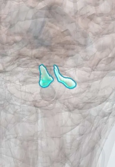
The cerebellar peduncles are the stalks of nerve fibres joining the cerebellum to the brainstem; they carry signals into and out of the cerebellum.
Official name: Superior cerebellar peduncle
Opens in the Durgical app, no sign-up needed.
What it is
The stalks of fibres joining the cerebellum to the brainstem.
What it does
Carry signals in and out of the cerebellum.
Key facts
- Where
- Superior (to the midbrain), middle (to the pons), inferior (to the medulla)
- Blood supply
- Superior cerebellar, AICA and PICA branches
- Function
- Middle: input from the cortex via the pons; inferior: input from the spinal cord and vestibular nuclei; superior: main output
Relations
- Above
- Superior peduncles climb to the midbrain on either side of the superior medullary velum, the roof of the upper fourth ventricle
- Lateral
- Middle peduncles on the sides of the pons; the trigeminal nerve emerges where pons and peduncle meet, with the facial and vestibulocochlear nerves just below
- Below
- Inferior peduncles on the back and side of the medulla oblongata, above the gracile and cuneate tubercles
- Medial
- Fourth ventricle, whose side walls they form
How to find it
In dissection they appear as three cut stalks when the cerebellum is lifted off the brainstem: the superior toward the midbrain, the middle (the largest) from the pons and the inferior from the back of the medulla. On axial MRI the middle peduncles are the broad bands joining the pons to each cerebellar hemisphere.
On imaging
On axial T2 MRI the middle peduncles are the thick bands joining the pons to each cerebellar hemisphere. Symmetric bright signal there suggests multiple system atrophy (cerebellar type) or fragile X-associated tremor/ataxia syndrome; diffusion tractography can trace all three peduncles.
Clinical notes
- Lateral medullary (Wallenberg) syndrome includes the inferior peduncle, adding same-side limb ataxia
- Multiple sclerosis plaques in the peduncles cause ataxia and tremor
- An AICA infarct hits the middle peduncle and the lateral pons, with same-side ataxia, facial weakness and hearing loss
Development
The superior peduncles and the superior medullary velum come from the isthmus, the narrow junction of midbrain and hindbrain. In Joubert syndrome the superior peduncles are thick, long and fail to cross, which with a small vermis gives the molar tooth sign on axial MRI.
Memory aid
Superior Sends out; Middle and Inferior bring In.
Related conditions
Test yourself
Think of the answer, then tap the question to see it.
Which cerebellar peduncle is mainly output, and where does it go?
The superior peduncle, from the dentate nucleus; it crosses in the lower midbrain and ends in the red nucleus and the ventrolateral thalamus.
What does the middle cerebellar peduncle carry?
Purely input: crossed fibres from the pontine nuclei (the cortico-ponto-cerebellar route); it is the largest peduncle.
Which inputs travel in the inferior cerebellar peduncle?
From the spinal cord (dorsal spinocerebellar, cuneocerebellar), the opposite inferior olive and the vestibular system.
Educational information, not medical advice. For any symptom, see a health professional.
Its names
- Official Latin (TA2)
- pedunculus cerebellaris superior TA2 5846
- Official Spanish (DeCS)
- Pedúnculos cerebelosos
- English (TA2)
- Superior cerebellar peduncle
- Also called
- brachium conjunctivum · pedunculus cerebellaris cranialis · pedunculus cerebellaris rostralis
Sources: Terminologia Anatomica (TA2); Neuroanatomy, Cerebellum; Neuroanatomy, Brainstem; Anatomy of the Human Body (20th ed.): Development of the Nervous System (Lea & Febiger (Gray's Anatomy, 1918), 1918) · adapted, Public domain; Anatomía humana (4.ª ed.), tomo 1, cap. 31: Configuración interna y sistematización del cerebelo.
Spotted a mistake?We are always improving for the community. Tell us what is wrong and our team will review it.Report a mistake
Representative model, for educational purposes only.