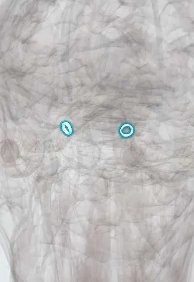
Common tendinous ring !
The common tendinous ring is a ring of tendon at the back of the eye socket where the four rectus eye muscles start; the optic nerve passes through it.
Opens in the Durgical app, no sign-up needed.
What it is
A ring of tendon at the back of the eye socket.
What it does
The four rectus muscles start here; the optic nerve passes through it.
Key facts
- Attaches
- Around the optic canal and the middle of the superior orbital fissure
- Function
- Origin of the four recti; II, ophthalmic artery, III, VI and the nasociliary nerve pass through it
Relations
- Contents
- Optic nerve and ophthalmic artery (from the optic canal); superior and inferior divisions of the oculomotor nerve (III); nasociliary nerve (V1); abducens nerve (VI); usually the sympathetic root of the ciliary ganglion
- Above
- Above and outside it: the lacrimal, frontal and trochlear nerves and the superior ophthalmic vein; the levator palpebrae superioris arises just above it
- In front
- The four recti fanning out around the optic nerve to form the muscle cone, with intraconal fat between them
- Behind
- Optic canal and superior orbital fissure, opening into the middle cranial fossa, with the cavernous sinus just behind
- Below
- The inferior ophthalmic vein, which often passes below the ring
How to find it
In dissection, the orbital roof and the levator are lifted and the four recti traced back to the apex, where they converge on a fibrous ring around the optic canal and the middle part of the superior orbital fissure.
On imaging
Thin-slice MRI with fat suppression and contrast shows the orbital apex and the nearby cavernous sinus: abnormal enhancing tissue suggests inflammation (such as Tolosa–Hunt syndrome), tumour or fungal infection; CT shows fractures and bone erosion of the apex.
Clinical notes
- Causes of orbital apex syndrome: fractures, tumours, inflammation and invasive fungal sinusitis (mucormycosis) in people with poorly controlled diabetes or immunosuppression
- Superior orbital fissure syndrome affects III, IV, V1 and VI but spares vision; once the optic nerve is involved it becomes an orbital apex syndrome
- Cavernous sinus lesions add the maxillary nerve (V2) and can spread to both sides; comparing these three syndromes localises the lesion
- A retrobulbar (intraconal) block places anaesthetic inside the muscle cone that starts at the ring; rare complications are retrobulbar haemorrhage, optic nerve injury and spread along the optic nerve sheath to the brainstem
- Swollen recti in thyroid eye disease crowd the apex at the ring and can compress the optic nerve (dysthyroid optic neuropathy), shown by falling colour vision and a relative afferent pupillary defect
Memory aid
Superior orbital fissure from top down: "Live Frankly To See Absolutely No Insult" (Lacrimal, Frontal, Trochlear outside the ring; Superior division of III, Abducens, Nasociliary, Inferior division of III inside it).
Test yourself
Think of the answer, then tap the question to see it.
Which structures pass through the common tendinous ring?
The optic nerve and ophthalmic artery, both divisions of the oculomotor nerve (III), the nasociliary nerve (V1) and the abducens nerve (VI).
Why may the superior oblique still move after a retrobulbar (intraconal) block?
The trochlear nerve passes outside the common tendinous ring, outside the muscle cone.
Why does optic neuritis hurt on eye movement?
The superior and medial recti attach to the optic nerve sheath at the common tendinous ring.
How does orbital apex syndrome differ from superior orbital fissure syndrome?
Both affect III, IV, V1 and VI; orbital apex syndrome also involves the optic nerve, so vision is lost.
Educational information, not medical advice. For any symptom, see a health professional.
Its names
- Official Latin (TA2)
- anulus tendineus communis TA2 2047
- Official Spanish (DeCS)
- Anillo tendinoso común
- English (TA2)
- Common tendinous ring
- Also called
- common anular tendon · anulus of Zinn
Sources: Terminologia Anatomica (TA2); Anatomy, Head and Neck, Eye Extraocular Muscles; Anatomy, Head and Neck, Orbit.
Spotted a mistake?We are always improving for the community. Tell us what is wrong and our team will review it.Report a mistake
Representative model, for educational purposes only.