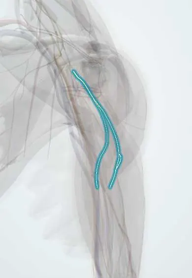
Thoracodorsal nerve !
The thoracodorsal nerve runs down the back wall of the armpit; it moves the latissimus dorsi, the broad muscle of the back.
Opens in the Durgical app, no sign-up needed.
What it is
A nerve from the posterior cord to the latissimus dorsi.
What it does
Powers latissimus dorsi.
Key facts
- Roots
- C6–C8, posterior cord
- Moves
- Latissimus dorsi
- Feeling from
- None
Relations
- Course
- Arises from the posterior cord between the upper and lower subscapular nerves, descends along the posterior axillary wall over subscapularis and teres major, joins the thoracodorsal artery and enters the deep surface of latissimus dorsi near its anterior border
- Behind
- Subscapularis and teres major on the posterior wall of the axilla
- In front
- Axillary fat and lymph nodes; the subscapular vessels
- Medial
- The long thoracic nerve on serratus anterior, across the axilla
How to find it
It cannot be felt. In axillary dissection it is seen running down the posterior axillary wall with the thoracodorsal vessels into latissimus dorsi, behind and lateral to the long thoracic nerve.
Examination
- Cough test: the examiner grips the posterior axillary folds while the person coughs; a fold that does not tense indicates latissimus dorsi weakness
- Resisted adduction and extension of the arm from abduction (pulling down against resistance), with the posterior axillary fold palpated
On imaging
Not usually seen on its own. It runs with the thoracodorsal artery and vein on the deep surface of latissimus dorsi, and these vessels are the landmark on ultrasound or contrast CT, for example when planning a latissimus dorsi flap. After nerve injury, MRI shows oedema of latissimus dorsi and later fatty atrophy; electromyography confirms the denervation.
Clinical notes
- Injury in axillary surgery weakens adduction and internal rotation, a loss that is often well tolerated in daily life
- In latissimus dorsi flap reconstruction the nerve may be divided to reduce visible muscle twitching, or kept to preserve muscle bulk
- A donor nerve for transfers, for example to the long thoracic nerve or to restore shoulder and elbow function
Anatomical variants
- Its origin varies: it may arise from the axillary or radial nerve rather than from the cord itself, and the roots feeding it differ between people
- It usually divides into a lateral and a medial branch before entering the muscle, which allows split-muscle flaps
Development
Latissimus dorsi is a limb muscle that spreads back over the trunk during development and keeps its posterior cord nerve; this long nerve and vessel pedicle is what lets the muscle be moved as a flap.
Test yourself
Think of the answer, then tap the question to see it.
Which muscle does the thoracodorsal nerve supply, and along which axillary wall does it run?
Latissimus dorsi; it runs down the posterior wall of the axilla with the thoracodorsal vessels.
What is the role of the thoracodorsal nerve and vessels in breast reconstruction?
They form the pedicle of the latissimus dorsi flap.
What is the cough test for the thoracodorsal nerve?
The examiner grips the posterior axillary folds while the person coughs; a fold that does not tense indicates latissimus dorsi weakness.
Educational information, not medical advice. For any symptom, see a health professional.
Its names
- Official Latin (TA2)
- nervus thoracodorsalis TA2 6430
- Official Spanish (DeCS)
- Nervio toracodorsal
- English (TA2)
- Thoracodorsal nerve
- Also called
- middle subscapular nerve
Related
Sources: Terminologia Anatomica (TA2); Anatomy, Thorax, Thoracodorsal Nerves; Anatomy of the Human Body (20th ed.): Development of the Muscles (Lea & Febiger (Gray's Anatomy, 1918), 1918) · adapted, Public domain.
Spotted a mistake?We are always improving for the community. Tell us what is wrong and our team will review it.Report a mistake
Representative model, for educational purposes only.