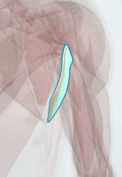
Teres major !
The teres major runs from the lower shoulder blade to the humerus; with the latissimus dorsi it pulls the arm down and back and turns it inward.
Opens in the Durgical app, no sign-up needed.
What it is
A thick muscle from the lower corner of the shoulder blade to the front of the humerus.
What it does
Pulls the arm down and back and turns it inward, with the latissimus dorsi.
Key facts
- Origin
- Posterior surface of the inferior angle of the scapula
- Insertion
- Medial lip of the intertubercular sulcus
- Action
- Adducts, extends and medially rotates the arm
- Nerve
- Lower subscapular nerve (C5–C7)
- Blood supply
- Circumflex scapular artery, thoracodorsal artery
Relations
- Above
- Teres minor, separated laterally by the triceps (long head): the quadrangular space lies lateral to the long head, the triangular space medial to it
- Below
- The triangular interval, below it between the triceps (long head) and the humerus, where the radial nerve and deep brachial artery reach the back of the arm
- In front
- Latissimus dorsi, whose tendon wraps round its lower border to insert in front of it; the radial nerve and brachial artery cross in front of both tendons
- Lateral
- Medial lip of the intertubercular groove, beside the latissimus tendon
How to find it
With the arm slightly abducted and pressed backward and inward against resistance, it forms the thick rounded mass of the lower posterior axillary fold, running from the inferior angle of the scapula toward the armpit.
Examination
- Manual muscle test (prone, hand on the lower back, examiner pushing the elbow toward the floor): weakness of extension and adduction points to the lower subscapular nerve or the C5–C6 roots
On imaging
On sagittal oblique MRI it lies below the teres minor, with the long head of the triceps passing between them; its tendon inserts on the medial lip of the bicipital groove beside the latissimus dorsi tendon. Isolated tears, mostly in throwers, show on MRI as a tendon gap with oedema. On angiography its lower border marks where the axillary artery becomes the brachial.
Clinical notes
- Its lower border is the landmark used to name and expose the vessel: above it axillary artery, below it brachial artery
- Transfer of the latissimus dorsi and teres major to the back of the humerus is used to give external rotation in brachial plexus birth palsy
- The spaces it bounds are landmarks in posterior shoulder surgery and in quadrangular space syndrome
Anatomical variants
- Variants are rare and mostly at the insertion, where it may fuse with the latissimus dorsi tendon
Development
Develops from the dorsal muscle mass together with latissimus dorsi, which explains why both are supplied from the posterior cord and insert side by side on the medial lip and floor of the intertubercular groove.
Memory aid
'A lady between two majors': the latissimus dorsi inserts on the floor of the groove between pectoralis major (lateral lip) and teres major (medial lip).
Test yourself
Think of the answer, then tap the question to see it.
Where does the axillary artery become the brachial artery?
At the lower border of the teres major.
Is the teres major part of the rotator cuff, and what nerve supplies it?
No; it works with the latissimus dorsi and is supplied by the lower subscapular nerve (C5–C7).
What passes through the triangular interval below the teres major?
The radial nerve and the deep brachial artery, on their way to the back of the arm.
Where does the teres major insert, and what are its actions?
On the medial lip of the intertubercular sulcus; it adducts, extends and medially rotates the arm.
Educational information, not medical advice. For any symptom, see a health professional.
Its names
- Official Latin (TA2)
- musculus teres major TA2 2462
- Official Spanish (DeCS)
- Músculo redondo mayor
- English (TA2)
- Teres major
Related
Sources: Terminologia Anatomica (TA2); Anatomy, Shoulder and Upper Limb, Teres Major Muscle.
Spotted a mistake?We are always improving for the community. Tell us what is wrong and our team will review it.Report a mistake
Representative model, for educational purposes only.