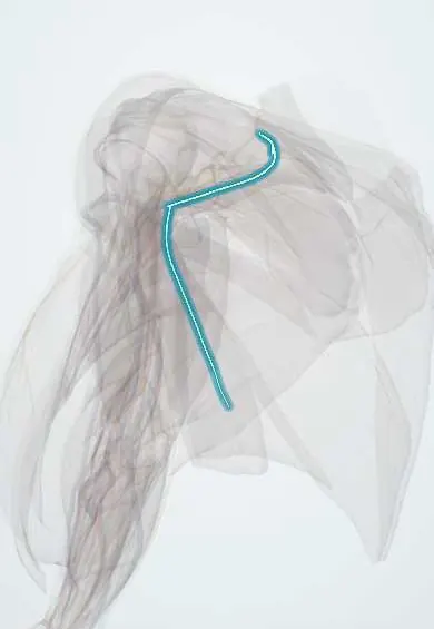
Long thoracic nerve !
The long thoracic nerve runs down the side of the chest wall; it moves the serratus anterior, and damage makes the shoulder blade stick out (winging).
Opens in the Durgical app, no sign-up needed.
What it is
A long nerve down the side of the chest to serratus anterior; injury (heavy backpacks, sports, surgery) causes a winged scapula.
What it does
Powers serratus anterior; damage makes the shoulder blade stick out (winging).
Key facts
- Roots
- C5–C7 roots (some texts: C6–C7)
- Moves
- Serratus anterior (injury = winged scapula)
- Feeling from
- None
Relations
- Course
- The C5 and C6 roots pierce the middle scalene and are joined by C7; the nerve descends behind the brachial plexus and the first part of the axillary artery, crosses the first rib into the axilla and runs down the outer surface of serratus anterior near the midaxillary line, supplying each digitation
- Deep
- Serratus anterior, on whose surface it lies
- In front
- The lateral thoracic artery, which runs down in front of it
- Behind
- The thoracodorsal nerve and vessels on the posterior wall of the axilla
- Superficial
- Axillary fat and lymph nodes and the skin of the lateral chest wall
How to find it
Not palpable. In axillary surgery it is identified on the chest wall under a thin fascial layer over serratus anterior, near the midaxillary line, in front of the thoracodorsal bundle; both nerves are preserved.
Examination
- Wall push-up: pushing against a wall with straight arms makes the medial border and inferior angle of the scapula lift off the chest; positive = serratus anterior weakness
- Forward elevation of the arm is limited and painful; pressing the scapula against the ribs by hand (scapular assistance) improves it
On imaging
Not seen on routine imaging; the diagnosis is clinical (medial winging when pushing on a wall) and confirmed by electromyography of serratus anterior. MRI of the chest wall can show oedema of serratus anterior early and fatty atrophy later. MRI of the cervical spine or brachial plexus excludes a root or plexus lesion, and ultrasound can follow the nerve on the serratus in the mid-axillary line.
Common problems
- Winged scapula: the inner edge of the shoulder blade lifts off the ribs when the long thoracic nerve is injured.
- Weakness makes overhead reaching and pushing hard and can strain the shoulder.
How it is diagnosed
Wall push-up test shows the winging. EMG checks the long thoracic nerve.
Treatment
- Physical therapy; most nerve stretch injuries recover over months.
- Muscle or tendon transfer surgery if the nerve does not recover.
Clinical notes
- Injured in axillary lymph node dissection, mastectomy, first rib resection, chest drain insertion and lateral thoracotomy
- Non-surgical causes: heavy loads on the shoulder strap (rucksack palsy), repeated overhead sport and neuralgic amyotrophy
- Most stretch palsies recover slowly over months; persistent winging may be treated by transferring the sternal head of pectoralis major to the scapula
Anatomical variants
- A small C8 contribution is reported in about one person in ten, and the C7 root may be missing
Development
Serratus anterior is not a body-wall muscle like the intercostals: it develops with the limb musculature and spreads onto the chest wall, which is why its nerve comes from the plexus roots (C5–C7) and not from intercostal nerves.
Memory aid
'C5, 6, 7, wings of heaven': the long thoracic nerve (C5–C7) keeps the scapula on the chest; injury gives a winged scapula. 'SALT': Serratus Anterior, Long Thoracic.
Related conditions
Test yourself
Think of the answer, then tap the question to see it.
Which muscle does the long thoracic nerve supply, and which roots form it?
Serratus anterior; C5–C7 roots.
What scapular sign follows long thoracic nerve palsy, and how is it shown?
Medial winging: the medial border and inferior angle lift off the chest wall, best seen with a wall push-up.
How does winging from spinal accessory nerve palsy differ from long thoracic nerve palsy?
Trapezius palsy gives lateral winging with a drooping shoulder; serratus anterior palsy gives medial winging.
In which operations is the long thoracic nerve at risk?
Axillary lymph node dissection, mastectomy, first rib resection, chest drain insertion and lateral thoracotomy; it lies on serratus anterior on the medial axillary wall.
Educational information, not medical advice. For any symptom, see a health professional.
Its names
- Official Latin (TA2)
- nervus thoracicus longus TA2 6410
- Official Spanish (DeCS)
- Nervio torácico largo
- English (TA2)
- Long thoracic nerve
Related
Sources: Terminologia Anatomica (TA2); Anatomy, Thorax, Long Thoracic Nerve; Anatomy of the Human Body (20th ed.): Development of the Muscles (Lea & Febiger (Gray's Anatomy, 1918), 1918) · adapted, Public domain.
Spotted a mistake?We are always improving for the community. Tell us what is wrong and our team will review it.Report a mistake
Representative model, for educational purposes only.