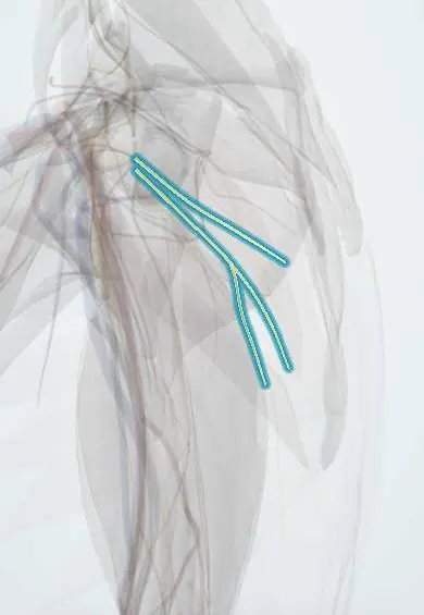
Subscapular nerves !
The subscapular nerves are two short nerves in the back of the armpit; they move the subscapularis and teres major.
Official name: Upper and lower subscapular nerves
Opens in the Durgical app, no sign-up needed.
What it is
Two short nerves from the posterior cord to the front of the shoulder blade.
What it does
Power subscapularis and teres major.
Key facts
- Roots
- C5–C6, posterior cord
- Moves
- Upper: subscapularis; lower: subscapularis and teres major
- Feeling from
- None
Relations
- Course
- Arising from the posterior cord in the axilla, the upper nerve enters the upper part of subscapularis high in the axilla; the lower nerve descends beside the subscapular vessels to the lower part of subscapularis and on to teres major
- In front
- The axillary artery and its subscapular branch
- Behind
- Subscapularis on the costal surface of the scapula
- Lateral
- The axillary nerve, which crosses the lower border of subscapularis toward the quadrangular space
How to find it
Not palpable. They enter subscapularis medial to the coracoid process, so in an anterior (deltopectoral) approach the muscle can be divided or released near the humerus without denervating it; dissection far medially on the muscle puts them at risk.
Examination
- Lift-off test (Gerber): with the back of the hand on the lower back, lifting it away is weak or impossible when subscapularis is weak
- Belly-press test: pressing the palm into the abdomen with the elbow held forward; the elbow dropping back, with the wrist flexing to compensate, indicates subscapularis weakness
- Bear-hug test: holding the hand on the opposite shoulder against an upward pull; failure indicates weakness of the upper subscapularis
On imaging
Not seen on routine imaging; their state is judged on the muscles they supply. On shoulder MRI, denervation of subscapularis or teres major shows as muscle oedema early and fatty atrophy later. Ultrasound and MRI also show tears of the subscapularis tendon, which must be told apart from weakness caused by nerve injury.
Clinical notes
- Rarely injured alone; they can be damaged in posterior cord injuries, axillary surgery and extensive medial release of subscapularis
- Weak internal rotation with an intact subscapularis tendon on imaging suggests a nerve lesion rather than a tear
Anatomical variants
- The lower subscapular nerve may arise from the axillary nerve, and either nerve may be doubled
- Prefixed or postfixed plexuses change their root content
Development
Subscapularis and teres major come from the dorsal muscle mass, so their nerves arise from the posterior cord although the muscles face forward into the axilla.
Test yourself
Think of the answer, then tap the question to see it.
Which muscles do the upper and lower subscapular nerves supply?
Upper: subscapularis; lower: subscapularis and teres major.
Which nerve arises between the upper and lower subscapular nerves from the posterior cord?
The thoracodorsal nerve, also called the middle subscapular nerve.
Which tests examine subscapularis, supplied by the subscapular nerves?
The lift-off (Gerber), belly-press and bear-hug tests.
Educational information, not medical advice. For any symptom, see a health professional.
Its names
- Official Latin (TA2)
- nervi subscapulares TA2 6427
- Official Spanish (DeCS)
- Nervios subescapulares superior e inferior
- English (TA2)
- Upper and lower subscapular nerves
Related
Sources: Terminologia Anatomica (TA2); Anatomy, Shoulder and Upper Limb, Nerves; Anatomy, Shoulder and Upper Limb, Brachial Plexus; Anatomy of the Human Body (20th ed.): The Branchial Region (Lea & Febiger (Gray's Anatomy, 1918), 1918) · adapted, Public domain.
Spotted a mistake?We are always improving for the community. Tell us what is wrong and our team will review it.Report a mistake
Representative model, for educational purposes only.