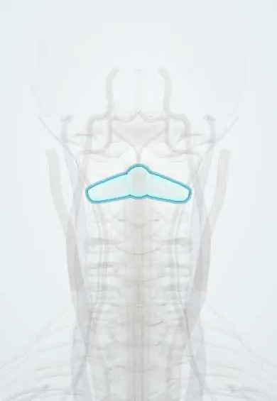
Atlas–axis joints !
The atlantoaxial joints are the three joints between C1 and C2; they provide about half of all neck rotation, as in shaking the head "no".
Official name: Median and lateral atlanto-axial joints
Opens in the Durgical app, no sign-up needed.
What it is
The three joints between the atlas and the axis: two flat side joints and a pivot joint around the dens.
What it does
Allow about half of all neck rotation (around 40° to each side) when you shake your head 'no'.
Key facts
- Where
- Median: dens with anterior arch of atlas and transverse ligament; two lateral: atlas lateral masses on C2 superior facets (C1–C2)
- Attaches
- Cruciform (transverse) and alar ligaments; tectorial membrane; joint capsules; no intervertebral disc at C1–C2
- Function
- Median is a synovial pivot, lateral are plane joints; head rotation about 40° each side (some texts: about 30°), about half of all neck rotation
Relations
- In front
- Anterior arch of the atlas in front of the dens, the anterior atlantoaxial membrane and longus colli; further forward, the retropharyngeal space and the pharynx
- Behind
- Median joint: the transverse ligament of the atlas, tectorial membrane, dura and spinal cord; lateral joints: the C2 root and ganglion behind each capsule
- Lateral
- The vertebral artery rising through the transverse foramina of C2 and C1, just outside the lateral joints
- Below
- The C2–C3 disc and facet joints: the first true intervertebral disc of the spine
Examination
- Cervical flexion-rotation test: the neck is fully flexed to limit movement at lower levels and the head is turned to each side; clearly less rotation on one side suggests a C1–C2 source of cervicogenic headache
On imaging
The open-mouth (odontoid) view shows the C1 lateral masses sitting on C2: overhang beyond C2 suggests a Jefferson fracture, and unequal spaces beside the dens may be rotation or injury. Lateral flexion-extension views measure the ADI. Dynamic CT with the head turned to each side shows rotatory fixation.
Common problems
- Neck pain and headaches from arthritis or after whiplash.
- Bone spurs that narrow the opening for a nerve root.
How it is diagnosed
There is no single sign. CT or MRI show arthritis; a numbing injection of the nerves to the joint (medial branch block) shows whether the joint is the pain source.
Treatment
- Physical therapy, medicine, injections; radiofrequency ablation for lasting facet pain.
Clinical notes
- Atlantoaxial instability in rheumatoid arthritis (synovitis of the median joint erodes the dens and its ligaments) and in Down syndrome (ligament laxity); the cord is at risk with neck flexion
- Posterior C1–C2 fusion (transarticular screws, or C1 lateral mass and C2 pars or pedicle screws) crosses these joints; the vertebral artery is the structure at risk
- After C1–C2 fusion, roughly half of neck rotation is lost
Anatomical variants
- Os odontoideum: a smooth, rounded ossicle above a short dens, separated by a gap with corticated edges; it can make C1–C2 unstable, and whether it is developmental or the result of an old injury is debated
Development
The dens forms from its own ossification centres and is joined to the body of C2 by a cartilage plate (subdental synchondrosis) that usually fuses in early childhood; until then the line can be mistaken for a fracture.
Memory aid
The atlas says yes (C0–C1 nods), the axis says no (C1–C2 turns).
Related conditions
Test yourself
Think of the answer, then tap the question to see it.
How much neck rotation occurs at the atlantoaxial joints?
About 40° to each side, roughly half of all neck rotation; it is lost after a C1–C2 fusion.
What is the normal atlantodental interval (ADI) on a lateral view, and what does widening mean?
Up to about 3 mm in adults and 5 mm in children; a wider gap means the transverse ligament of the atlas is failing.
Which nerve root lies directly behind the lateral atlantoaxial joint?
The C2 root and its ganglion, not in a foramen; arthritis of this joint causes suboccipital pain and headache.
What is Grisel syndrome?
Atlantoaxial rotatory subluxation in a child after a throat or neck infection, giving a fixed, tilted and turned head.
Which structure is at risk during posterior C1–C2 fusion with screws across the atlantoaxial joints?
The vertebral artery, which rises through the transverse foramina of C2 and C1 just outside the lateral joints.
Educational information, not medical advice. For any symptom, see a health professional.
Its names
- Official Latin (TA2)
- articulatio atlantoaxialis mediana; articulatio atlantoaxialis lateralis TA2 1694
- Official Spanish (DeCS)
- Articulación atlantoaxoidea DeCS D001268
- English (TA2)
- Median and lateral atlanto-axial joints
- Also called
- Atlanto-Axial Joint · articulatio atlantoepistrophica · articuli atlantodentales
Related
Sources: Terminologia Anatomica (TA2); DeCS; Anatomy, Head and Neck: Atlantoaxial Joint.
Spotted a mistake?We are always improving for the community. Tell us what is wrong and our team will review it.Report a mistake
Representative model, for educational purposes only.