Conditions · Pediatrics and neonatology
Achondroplasia !
Achondroplasia is the most common inherited form of short-limbed dwarfism. A change in the FGFR3 gene slows bone growth; intelligence is normal.
Updated 2026-11-01
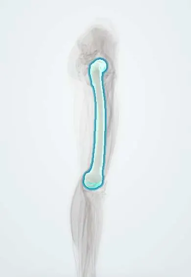
Structures involved
Tap one to see what it is and open it in 3D.
Why it happens
- Mendelian inheritance: autosomal dominant, recessive and X-linked
- How a single gene causes disease: enzymes, receptors and structural proteins
- Receptor tyrosine kinases
- Ossification, growth plate and bone remodeling
- Cartilage: hyaline, elastic, fibrocartilage
What it is
A genetic skeletal dysplasia in which growth of bone from cartilage (endochondral ossification) is impaired, producing disproportionate short stature with short proximal limbs, a large head and a normal-length trunk.
Who gets it
Occurs in roughly 1 in 15,000 to 40,000 births worldwide, in all populations and both sexes. About four in five cases are new mutations in children of average-height parents; paternal age over 35 raises the chance. In Latin America it is usually recognised at birth or on prenatal ultrasound in the third trimester.
Causes
- Gain-of-function variant of the FGFR3 gene (fibroblast growth factor receptor 3), almost always the same point change (G380R)
- Autosomal dominant: one altered copy is enough
- About 80% are de novo mutations; the rest are inherited from an affected parent
- Two altered copies (both parents affected) cause homozygous achondroplasia, usually lethal in infancy
Risk factors
- An affected parent (50% risk for each child)
- Advanced paternal age for de novo cases
- Two affected parents (25% risk of the lethal homozygous form)
Pathophysiology
The overactive FGFR3 receptor puts a brake on chondrocyte proliferation in the growth plates, so long bones grow slowly in length while periosteal (membranous) bone growth in width is normal. The skull base and vertebrae, which form from cartilage, stay small: a narrow foramen magnum can compress the cervicomedullary junction in infancy, and short pedicles narrow the lumbar spinal canal, causing stenosis in adulthood.
Classification
- Heterozygous achondroplasia (typical form)
- Homozygous achondroplasia (severe, usually lethal)
- Same FGFR3 spectrum: hypochondroplasia (milder) and thanatophoric dysplasia (lethal)
Symptoms and signs
- Disproportionate short stature with rhizomelia (short upper arms and thighs)
- Macrocephaly with frontal bossing and midface hypoplasia with a depressed nasal bridge
- Trident hand (gap between the middle and ring fingers) and limited elbow extension
- Infant hypotonia with delayed motor milestones; thoracolumbar kyphosis in infancy
- Exaggerated lumbar lordosis and bowed legs (genu varum) in childhood
- Recurrent otitis media and conductive hearing loss; normal intelligence
Diagnosis
- Clinical
- Usually recognised at birth from the characteristic proportions, head shape and hands. Measure length, head circumference and weight on achondroplasia-specific growth charts, and examine tone, reflexes and the spine.
- Tests
- Molecular testing for the FGFR3 variant confirms the diagnosis when features are atypical or prenatally. Polysomnography screens for sleep apnoea; regular hearing assessment.
- Imaging
- Skeletal survey: short tubular bones with flared metaphyses, narrowing of the lumbar interpedicular distance going down the spine, squared iliac wings and flat acetabular roofs. MRI or CT of the craniocervical junction to assess foramen magnum stenosis in infants; prenatal ultrasound shows short femurs in the third trimester.
Differential diagnosis
- Hypochondroplasia (milder FGFR3 dysplasia)
- Thanatophoric dysplasia (lethal, more severe shortening)
- Pseudoachondroplasia (normal head and face, onset after age 2)
- Other causes of short stature (growth hormone deficiency, hypothyroidism, rickets)
Goal of treatment
Detect and treat neurological, respiratory and orthopaedic complications early, support development, hearing and healthy weight, and promote independence through lifelong multidisciplinary follow-up.
Treatment
- Medical
- A C-type natriuretic peptide analogue (vosoritide), which counteracts FGFR3 signalling, can increase growth velocity in children with open growth plates; availability varies by country.
- Surgical
- Decompression of the foramen magnum for symptomatic cervicomedullary compression; shunting for progressive hydrocephalus; lumbar laminectomy for spinal stenosis; osteotomy for severe bowing.
- Procedure
- Ear ventilation tubes for recurrent otitis media and adenotonsillectomy for obstructive sleep apnoea.
- Follow-up
- Specific growth charts, neurological and sleep assessment in infancy, hearing checks, weight control and spine review; safe infant positioning with head and neck support and avoidance of unsupported sitting early on.
Complications
- Cervicomedullary compression and sudden infant death
- Hydrocephalus
- Sleep apnoea
- Lumbar spinal stenosis with neurogenic claudication
- Recurrent otitis media and conductive hearing loss
- Obesity, kyphosis and genu varum
Prognosis
Intelligence and life expectancy are close to normal for most people, although infant mortality is higher from craniocervical compression. Average untreated adult height is about 131 cm in men and 124 cm in women. Most adults live independently; back and leg symptoms from spinal stenosis are common.
Prevention
- Genetic counselling for affected adults and families
- Prenatal or preimplantation genetic testing when a parent is affected
Test yourself
Think of the answer, then tap the question to see it.
Which gene and mechanism cause achondroplasia?
A gain-of-function mutation of FGFR3, which inhibits chondrocyte proliferation in the growth plate.
What is the inheritance pattern, and how do most cases arise?
Autosomal dominant; about 80% are de novo mutations, linked to advanced paternal age.
Which type of bone formation is impaired, and which is spared?
Endochondral ossification is impaired (long bones, skull base); membranous ossification is normal (skull vault, bone width).
What life-threatening complication is screened for in infants with achondroplasia?
Foramen magnum stenosis with cervicomedullary compression, which can cause central apnoea and sudden death.
What typical spinal problem appears in adults with achondroplasia?
Lumbar spinal stenosis from short pedicles, causing neurogenic claudication.
Related conditions
- Lumbar spinal stenosis
- Obstructive sleep apnoea
- Hydrocephalus
- Acute otitis media
- Osteogenesis imperfecta
Educational information, not medical advice. For any symptom, see a health professional.
Sources: Achondroplasia: MedlinePlus Genetics (MedlinePlus, US National Library of Medicine, 2026).
Spotted a mistake?We are always improving for the community. Tell us what is wrong and our team will review it.Report a mistake
Representative model, for educational purposes only.