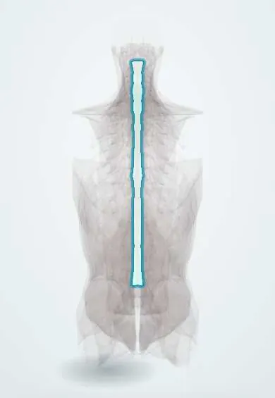
Lumbar spinal stenosis !
Lumbar spinal stenosis is narrowing of the lower spinal canal that squeezes the nerves; it causes leg pain on walking that eases on sitting.
Updated 2026-10-29
Structures involved
Tap one to see what it is and open it in 3D.
- Ligamentum flavum
- Low-back facet joints
- Cauda equina
- Dura (dural sac)
- L3 vertebra
- L4 vertebra
- L5 vertebra
- L4–L5 disc
- L5 nerve root
In embryology
Why it happens
- How organs age: normal changes vs disease
- Synovial joints: capsule, synovial membrane and articular cartilage
What it is
Narrowing of the lumbar spinal canal, lateral recesses or foramina that compresses the cauda equina or nerve roots. Its typical symptom is neurogenic claudication: leg pain on walking or standing that eases on sitting or bending forward.
Who gets it
Mainly affects people over 50, and is a frequent reason for spine surgery in older adults. It becomes more common as the population ages, as in Colombia and the rest of Latin America.
Causes
- Degenerative (acquired): disc bulging, facet joint hypertrophy and thickening of the ligamentum flavum
- Degenerative spondylolisthesis
- Congenital narrow canal (for example short pedicles or achondroplasia)
- Less often: post-surgical scarring, trauma, Paget disease, epidural lipomatosis
Risk factors
- Age over 50
- Spondylosis and degenerative disc disease
- Degenerative spondylolisthesis, most often at L4–L5
- Congenitally narrow canal
- Previous lumbar surgery
Pathophysiology
Disc bulging in front and a thick ligamentum flavum and enlarged facet joints behind narrow the canal into a trefoil shape. Upright posture and extension narrow it further, causing venous congestion and ischaemia of the nerve roots during activity; flexion widens the canal and relieves symptoms.
Classification
- By location: central canal, lateral recess or foraminal
- By cause: congenital or acquired (mostly degenerative)
Symptoms and signs
- Neurogenic claudication: pain, heaviness, numbness or weakness in the buttocks and legs on walking or standing
- Relief on sitting or bending forward; walking uphill or leaning on a shopping cart is easier (shopping-cart sign)
- Low back pain, often long-standing
- Radicular pain when a lateral recess or foramen is narrowed
- Neurological examination often normal at rest
Diagnosis
- Clinical
- Diagnosis is clinical, by the history of neurogenic claudication relieved by flexion. Pedal pulses and walking tests help separate it from vascular claudication, which eases on simply standing still and gives cold feet with weak pulses.
- Tests
- Ankle-brachial index when peripheral artery disease is suspected. Electromyography in selected cases to separate it from peripheral neuropathy.
- Imaging
- MRI is the study of choice to show the site and degree of narrowing; CT myelography if MRI is not possible. Standing flexion and extension X-rays look for instability or spondylolisthesis. Imaging stenosis is common without symptoms and must match the clinical picture.
Differential diagnosis
- Vascular claudication (peripheral artery disease)
- Lumbar disc herniation
- Hip osteoarthritis
- Peripheral neuropathy
- Greater trochanteric pain syndrome
Goal of treatment
Reduce leg pain and improve walking distance and function; decompress the nerves when symptoms are severe or deficits progress.
Treatment
- Conservative
- Education, activity modification and flexion-based positions; walking aids or a stationary bicycle are often better tolerated.
- Medical
- NSAIDs and analgesics; drugs for neuropathic pain in some cases, with limited evidence.
- Procedure
- Epidural corticosteroid injections may give short-term relief of leg pain.
- Rehabilitation
- Physical therapy with flexion exercises, core strengthening and conditioning.
- Surgical
- Decompressive laminectomy for disabling symptoms that fail conservative care or for progressive deficits; fusion is added when there is instability or spondylolisthesis.
Complications
- Reduced walking distance, loss of independence and deconditioning
- Cauda equina syndrome (rare)
- Surgical complications: dural tear, infection, instability, restenosis
Prognosis
Many patients stay stable for years, and some improve without surgery; rapid worsening is uncommon. Decompression usually improves leg pain and walking more than back pain.
Prevention
- Regular physical activity and trunk strengthening
- Healthy weight
Test yourself
Think of the answer, then tap the question to see it.
What is the shopping-cart sign in lumbar spinal stenosis?
Walking is easier leaning forward on a cart, because lumbar flexion widens the canal and relieves neurogenic claudication.
How is neurogenic claudication told apart from vascular claudication?
Neurogenic eases on sitting or bending forward, not on just standing still, and pulses are normal; vascular eases on standing still and pulses are weak.
Which three structures narrow the canal in degenerative lumbar stenosis?
The bulging disc in front, and the hypertrophied facet joints and thickened ligamentum flavum behind.
What is the imaging study of choice in lumbar spinal stenosis?
MRI of the lumbar spine; CT myelography when MRI cannot be done.
What operation is the standard surgical treatment for lumbar spinal stenosis?
Decompressive laminectomy, with fusion added if there is instability or spondylolisthesis.
Related conditions
- Sciatica (Lumbar Radiculopathy)
- Lumbar Disc Herniation
- Peripheral artery disease
- Cauda equina syndrome
- Mechanical low back pain
- Spondylolisthesis
- Abdominal aortic aneurysm
- Hip Osteoarthritis
- Greater Trochanteric Pain Syndrome
Educational information, not medical advice. For any symptom, see a health professional.
Sources: Lumbar Spinal Stenosis (StatPearls (NCBI Bookshelf), 2024).
Spotted a mistake?We are always improving for the community. Tell us what is wrong and our team will review it.Report a mistake
Representative model, for educational purposes only.