Adrenal incidentaloma !
An adrenal lump found by chance on a scan done for another reason. Most are harmless, but each needs hormone tests and a check that it is not cancer.
Updated 2026-11-01
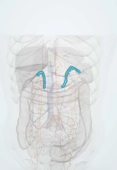
Structures involved
Tap one to see what it is and open it in 3D.
Why it happens
- Benign vs malignant tumours: the comparison table
- The HPA axis and its circadian rhythm
- Measuring hormones: stimulation and suppression tests
- Adrenal cortex zones and medulla
What it is
An adrenal mass, usually 1 cm or larger, discovered on imaging performed for a reason other than suspected adrenal disease. The two questions in every case are whether it secretes hormones and whether it is malignant.
Who gets it
Found in a few per cent of abdominal CT scans, and more often with age (over 5 to 7% of people older than 70). Its detection has risen with the wider use of CT in Colombia and the rest of Latin America. Most are benign non-functioning cortical adenomas.
Causes
- Non-functioning adrenocortical adenoma (most common)
- Adenoma with autonomous cortisol secretion (mild cortisol excess without full Cushing features)
- Aldosterone-producing adenoma and pheochromocytoma
- Adrenocortical carcinoma and metastases (lung, breast, kidney, melanoma, lymphoma)
- Myelolipoma, cyst, haemorrhage and granulomatous infection (tuberculosis, histoplasmosis)
Risk factors
- Older age
- Obesity, type 2 diabetes and hypertension (associated with adenomas)
- Known extra-adrenal cancer (raises the chance of metastasis)
Pathophysiology
Most incidentalomas are clonal benign growths of the adrenal cortex rich in intracellular lipid, which makes them low-density on unenhanced CT. Some secrete cortisol independently of ACTH; even modest excess raises the risk of hypertension, diabetes, osteoporosis and cardiovascular events. Malignant lesions are usually larger, lipid-poor and heterogeneous, and grow over time.
Classification
- By function: non-functioning, autonomous cortisol secretion, primary aldosteronism, pheochromocytoma
- By imaging: benign-appearing (homogeneous, 10 Hounsfield units or less on unenhanced CT) versus indeterminate
- By size: under 4 cm versus 4 cm or more, which raises concern for carcinoma
Symptoms and signs
- Usually none; found on a scan for abdominal pain, trauma or cancer staging
- Subtle features of cortisol excess: weight gain, hypertension, hyperglycaemia, bone loss
- Resistant hypertension or low potassium suggesting aldosterone excess
- Episodes of headache, sweating and palpitations suggesting pheochromocytoma
- Flank pain or weight loss with large or malignant masses
Diagnosis
- Clinical
- History and examination for hypertension, Cushing features, virilisation, adrenergic episodes and known cancer; review of the imaging that found the mass.
- Tests
- Every patient: 1 mg overnight dexamethasone suppression test for autonomous cortisol secretion and plasma or urine metanephrines for pheochromocytoma (metanephrines can be omitted if the mass is clearly lipid-rich adenoma on CT). Aldosterone-to-renin ratio if there is hypertension or low potassium. Sex steroid precursors if carcinoma is suspected. Biopsy is rarely needed and only after pheochromocytoma is excluded.
- Imaging
- Unenhanced CT is the key test: a homogeneous mass of 10 Hounsfield units or less is a benign adenoma. Indeterminate masses are assessed with contrast washout CT, chemical-shift MRI or FDG-PET.
Differential diagnosis
- Adrenocortical carcinoma
- Metastasis to the adrenal gland
- Pheochromocytoma
- Adrenal haemorrhage or cyst
- Pseudo-adrenal masses: upper pole renal lesion, splenic lobule, vessel or gastric diverticulum
Goal of treatment
Identify the few masses that are malignant or hormonally active and treat them, while sparing the many benign non-functioning adenomas from unnecessary surgery and repeated tests.
Treatment
- Surgical
- Adrenalectomy, usually laparoscopic, for functioning tumours (pheochromocytoma, aldosterone-producing adenoma, overt Cushing syndrome), suspected carcinoma or indeterminate masses that grow; selected cases of autonomous cortisol secretion with related disease.
- Medical
- Preoperative alpha-blockade for pheochromocytoma; perioperative glucocorticoid cover after removal of a cortisol-secreting adenoma because the other adrenal is suppressed. Treatment of hypertension, diabetes and bone loss in non-operated patients with mild cortisol excess.
- Follow-up
- Benign-appearing small non-functioning adenomas need no further imaging. Indeterminate masses are re-imaged at about 6 to 12 months. Mild cortisol excess is reviewed for its metabolic complications.
Complications
- Missed pheochromocytoma causing a hypertensive crisis during surgery or biopsy
- Cardiometabolic disease from autonomous cortisol secretion
- Adrenal insufficiency after removal of a cortisol-secreting adenoma
- Delayed diagnosis of adrenocortical carcinoma or metastasis
Prognosis
Excellent for benign non-functioning adenomas, which rarely grow or become active. Prognosis is driven by the underlying lesion when it is a carcinoma or metastasis.
Test yourself
Think of the answer, then tap the question to see it.
Which two questions must be answered for every adrenal incidentaloma?
Is it hormonally active, and is it malignant?
Which unenhanced CT finding confirms a benign lipid-rich adenoma?
A homogeneous mass with attenuation of 10 Hounsfield units or less.
Which screening test detects autonomous cortisol secretion?
The 1 mg overnight dexamethasone suppression test.
Why must pheochromocytoma be excluded before adrenal biopsy or surgery?
Manipulation can release catecholamines and trigger a hypertensive crisis.
Related conditions
- Cushing syndrome
- Phaeochromocytoma
- Primary hyperaldosteronism (Conn syndrome)
- Adrenal insufficiency
- Adrenal crisis
- Arterial hypertension
- Type 2 diabetes mellitus
- Osteoporosis
- Obesity
- Lung cancer
- Tuberculosis
- Histoplasmosis
Educational information, not medical advice. For any symptom, see a health professional.
Sources: ESE Clinical Practice Guideline on the management of Adrenal Incidentalomas, in collaboration with the European Network for the Study of Adrenal Tumors (European Society of Endocrinology, 2023); Nonfunctional Adrenal Masses - Endocrinology - MSD Manual Professional Edition (MSD Manual Professional Edition, 2025).
Spotted a mistake?We are always improving for the community. Tell us what is wrong and our team will review it.Report a mistake
Representative model, for educational purposes only.