Conditions · Nephrology and urology
Atheroembolic renal disease (cholesterol crystal embolism) !
Cholesterol crystals break off from plaques in the aorta and block tiny arteries in the kidneys, skin and gut, often after a catheter procedure.
Updated 2026-11-01
Structures involved
Tap one to see what it is and open it in 3D.
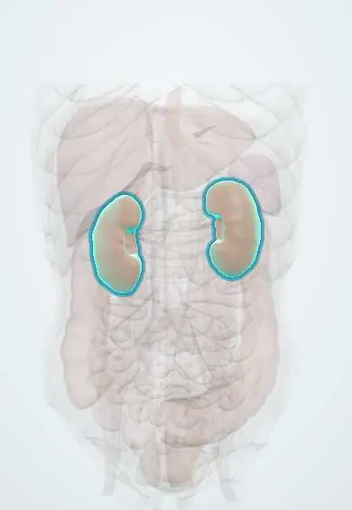
- Kidneys
- Left renal artery
- Toe arteries
- Right renal artery
- Vessels inside the kidney
- Abdominal aorta
- Superior mesenteric artery
- Skin
- Retina
Why it happens
- Granulomatous inflammation: caseating vs non-caseating, the causes to know
- Infarction: red vs white, and what decides the damage
- Fat, air and amniotic fluid embolism
- Systemic (arterial) thromboembolism
- Physiology of acute and chronic kidney failure
- Blood vessels: artery, vein and capillary layers
What it is
A form of systemic atheroembolism in which cholesterol crystals released from ulcerated atherosclerotic plaques of the aorta or large arteries lodge in small arteries. The kidneys are a frequent target, causing acute, subacute or chronic kidney injury, often together with skin lesions of the feet (blue toes, livedo reticularis).
Who gets it
Typical of older men with widespread atherosclerosis. It is probably underdiagnosed, since many cases are attributed to contrast nephropathy or to other causes of chronic kidney disease. With more coronary and peripheral angiography in Colombia and Latin America, it is a cause to consider in kidney injury after a procedure.
Causes
- Arterial catheterisation and angiography (coronary, aortic, renal), angioplasty and endovascular repair, the most common trigger
- Aortic or cardiac surgery that manipulates the aorta
- Anticoagulant or thrombolytic therapy, which may disrupt the plaque's protective thrombus
- Spontaneous plaque rupture without an obvious trigger
Risk factors
- Older age and male sex
- Advanced aortic atherosclerosis or abdominal aortic aneurysm
- Smoking, hypertension, diabetes and dyslipidaemia
- Known coronary, cerebrovascular or peripheral artery disease
- Pre-existing chronic kidney disease
Pathophysiology
Cholesterol crystals showered from an aortic plaque travel downstream and wedge in small arteries (arcuate and interlobular arteries in the kidney, small arteries of the skin, gut, retina and muscles). They provoke a foreign-body inflammatory reaction with intimal proliferation and fibrosis that gradually occludes the vessel, so kidney function often falls in steps over weeks rather than at once. Because the crystals dissolve during tissue processing, biopsies show empty, needle-shaped clefts.
Classification
- Acute (days after the trigger, often with many emboli)
- Subacute, with stepwise loss of kidney function over weeks (the most common pattern)
- Chronic, slowly progressive and clinically silent, mimicking nephrosclerosis
Symptoms and signs
- Rising creatinine and new or worsening hypertension days to weeks after a procedure
- Blue or purple toes with preserved foot pulses (blue toe syndrome)
- Livedo reticularis on the feet, legs, buttocks or abdomen
- Painful toe ulcers, nail-bed infarcts or small areas of gangrene
- Abdominal pain, gastrointestinal bleeding or pancreatitis from gut emboli
- Sudden painless visual loss in one eye from retinal emboli
- Fever, weight loss, muscle pain and malaise
Diagnosis
- Clinical
- Classic triad: a precipitating event (catheter procedure, vascular surgery or anticoagulation), acute or subacute kidney injury, and skin signs of the feet (blue toes with palpable pulses, livedo reticularis). Fundoscopy may show bright yellow cholesterol plaques at retinal artery branches (Hollenhorst plaques).
- Tests
- Rising creatinine; urinalysis with bland sediment or mild haematuria and modest proteinuria; transient eosinophilia (and sometimes eosinophiluria), low complement (C3) and raised ESR or CRP support the diagnosis but are not specific. Biopsy of kidney, skin or muscle showing biconvex needle-shaped clefts in small arteries confirms it; a skin biopsy of an affected area is less invasive.
- Imaging
- Not diagnostic. Ultrasound excludes obstruction and shows kidney size; CT or transoesophageal echo can show the extent of aortic plaque or aneurysm.
Differential diagnosis
- Contrast-associated acute kidney injury (earlier onset, usually recovers within days)
- Acute interstitial nephritis (drug-induced, also with eosinophilia)
- Systemic vasculitis such as polyarteritis nodosa or ANCA-associated vasculitis
- Renal artery thrombosis or thromboembolism from the heart
- Acute tubular necrosis from hypotension
- Calciphylaxis or warfarin skin necrosis for the skin lesions
Goal of treatment
There is no treatment that removes the crystals. The aims are to avoid further embolism, support kidney function, treat ischaemic organs and lower overall cardiovascular risk.
Treatment
- Acute
- Avoid further triggers: postpone non-essential catheter procedures or aortic surgery and review the need for anticoagulants or thrombolytics in each case.
- Medical
- Statins to stabilise plaques, aspirin as antiplatelet therapy, strict blood pressure control (often with ACE inhibitors or ARBs) and smoking cessation; glucocorticoids are sometimes used when inflammation is marked, with uncertain benefit.
- Conservative
- Supportive care: careful fluid and electrolyte management, nutrition, wound care of toe ulcers and pain control.
- Procedure
- Dialysis when kidney failure is severe; heparin-free sessions are often preferred. Some patients recover enough function to stop dialysis months later.
- Follow-up
- Nursing care: daily weight, fluid balance and urine output, serial creatinine and potassium, checking foot pulses and skin colour, and watching for abdominal pain or bleeding.
Complications
- Chronic kidney disease and end-stage kidney disease needing dialysis
- Accelerated or hard-to-control hypertension
- Bowel ischaemia, gastrointestinal bleeding and pancreatitis
- Toe gangrene and amputation
- Stroke and retinal artery occlusion
Prognosis
Guarded: many patients need dialysis and only some recover kidney function. Mortality is high, mostly from cardiovascular disease and multi-organ ischaemia rather than from kidney failure itself.
Prevention
- Careful indication of catheter procedures in people with severe aortic atherosclerosis; a radial approach may reduce aortic manipulation
- Statins and control of vascular risk factors
Test yourself
Think of the answer, then tap the question to see it.
What is the classic triad of atheroembolic renal disease?
A trigger such as an arterial catheter procedure, acute or subacute kidney injury, and skin signs of the feet such as blue toes or livedo reticularis.
Why can the toes be blue while the foot pulses are normal?
Cholesterol crystals block tiny distal arteries; the larger arteries that carry the pulses stay open.
Which laboratory findings support cholesterol embolism?
Transient eosinophilia, eosinophiluria, low complement (C3) and raised ESR, with a bland urine sediment.
What does the kidney biopsy show?
Empty biconvex needle-shaped clefts in arcuate and interlobular arteries, left where the cholesterol crystals dissolved during processing.
How does it differ in timing from contrast-associated kidney injury?
Contrast injury begins within a few days and usually recovers; atheroembolism often appears later and worsens stepwise over weeks.
Related conditions
- Acute kidney injury
- Chronic kidney disease
- Peripheral artery disease
- Abdominal aortic aneurysm
- Renovascular hypertension (renal artery stenosis)
- Polyarteritis nodosa
- Acute interstitial nephritis
- Acute mesenteric ischaemia
- Central retinal artery occlusion
Educational information, not medical advice. For any symptom, see a health professional.
Sources: Cholesterol Crystal Embolism and Chronic Kidney Disease (International Journal of Molecular Sciences, 2017) · adapted, CC BY 4.0.
Spotted a mistake?We are always improving for the community. Tell us what is wrong and our team will review it.Report a mistake
Representative model, for educational purposes only.